
Fetal Development
The actual embryo or fetal age (also known as conceptual age) is the time elapsed from fertilization of the egg near the time of ovulation. However, because most women do not know when ovulation occurred, but do know when their last period began, the time elapsed since the first day of the last normal menstrual period, the menstrual age, is used to determine the age of a pregnancy. The menstrual age is also known as the gestational age. Gestational age is conventionally expressed as completed weeks. Therefore, a 36 week, 6 day fetus is considered to be a 36 week fetus. [25]
Gestational Age Calculator (from Estimated Due Date)
The calculator below can be used to estimate the gestational age of a fetus on a given date. Enter the estimated due date, then enter the date you want to calculate the gestational age for and click Calculate.
| Structure / system | Critical period for major malformations Conceptional age (weeks after fertilization) |
Equivalent gestational age (LMP) (weeks) | Later vulnerability / notes |
|---|---|---|---|
| Central Nervous System (brain, spinal cord) |
~3–16 | ~5–18 | Functional effects (cognition, behavior, seizures, microcephaly) can occur from ~16 wks conceptional (~18 wks GA) through term. |
| Heart | ~3–6 | ~5–8 | Highest risk for major structural defects (e.g., conotruncal, septal). Minor structural/conduction issues still possible to ~8–10 wks conceptional (~10–12 wks GA). |
| Ears | ~4–9 | ~6–11 | Later (to ~16 wks conceptional / ~18 wks GA): mainly minor anomalies and sensorineural hearing loss risk. |
| Eyes | ~4–8 | ~6–10 | Functional and minor structural effects can extend from ~8 wks conceptional (~10 wks GA) to term. |
| Limbs (upper & lower) | ~4–5 | ~6–7 | High risk for reduction defects and major limb anomalies; smaller minor anomalies possible to ~8 wks conceptional (~10 wks GA). |
| Upper lip | ~5–6 | ~7–8 | Classic window for cleft lip / lip fusion defects. |
| Palate (secondary) | ~6–9 | ~8–11 | Cleft palate risk concentrated in this period. |
| Teeth | ~6–8 | ~8–10 | Disturbances of enamel / tooth development can continue from ~8 wks conceptional (~10 wks GA) into later fetal life. |
| External genitalia | ~7–9 | ~9–11 | Sex differentiation and external genital formation; virilization/undervirilization and some minor anomalies possible from ~9 wks conceptional (~11 wks GA) to term. |
| Urinary tract (kidneys, ureters, bladder, urethra) | ~4–8 | ~6–10 | Major structural anomalies arise primarily in this window; later in gestation, effects are more on growth and function rather than new major malformations. |
View critical-periods chart image
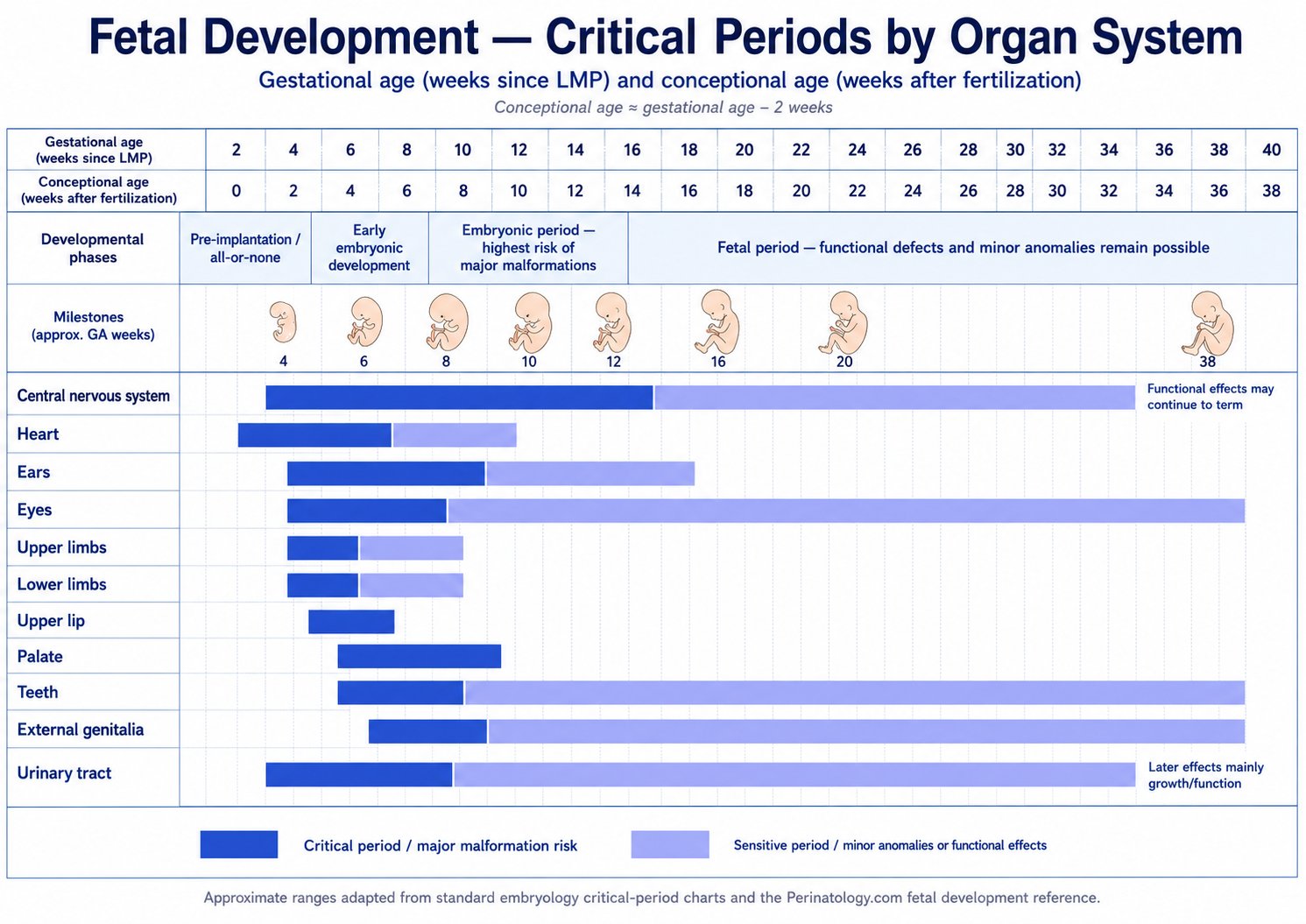
Conceptional age ≈ gestational age − 2 weeks. Ranges are approximate and adapted from standard critical-period charts (Moore, The Developing Human;).Sadler, Langman’s medical embryology;
Week-by-Week Fetal Development
First trimester of pregnancy
Less than 14 weeks 0 days (gestational age)Weeks 1 and 2 of pregnancy
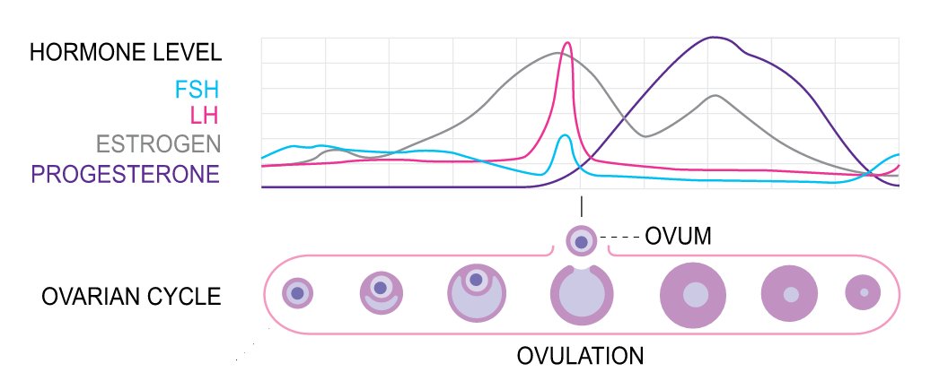
During the first two weeks after the last menstrual period, egg follicles mature in the ovaries under the stimulus of follicle-stimulating hormone (FSH), a hormone secreted by the pituitary gland in the brain.
High levels of the hormone estradiol, produced by the developing egg follicle, cause secretion of luteinizing hormone (LH), another hormone from the pituitary gland. LH triggers release of the egg from its follicle (ovulation).
For women with 28-day cycles, ovulation usually occurs on days 13 to 15.
Gestational age 3 weeks — embryonic age 1 week
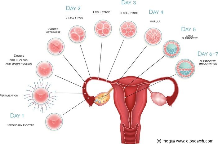
- During the third week, if fertilization occurs, the fertilized egg (zygote) begins producing the hormone human chorionic gonadotropin (hCG, the pregnancy hormone).
- hCG first becomes detectable in the mother's blood and urine between 6 and 14 days after fertilization (3 to 4 weeks gestational age).[7,8]
- During the 3rd week the sex of the fetus is determined by the father's sperm, and twins may be formed.
- Fatigue and swollen or tender breasts are sometimes the first signs of pregnancy.
Gestational age 4 weeks (0.9 months) — embryonic age 2 weeks
The embryo is approximately the size of a pinhead. Most pregnancy tests are positive at this time.
Gestational age 5 weeks (1.2 months) — embryonic age 3 weeks
- The brain, spine, and heart have begun to form.
- By the end of the week the heart will be pumping blood.
- Week 5 is the beginning of the embryonic period, which lasts from the 5th to the 10th week.
- It is during this critical period that many birth defects occur in the developing embryo. Most of these birth defects will have no known cause or will be due to a combination of factors (multifactorial).
Gestational age 6 weeks (1.4 months) — embryonic age 4 weeks

- The embryo is now about the size of a pea.
- The average crown–rump length is about 0.2 inches (0.5 cm).
- The eyes, nostrils, and arms are taking shape.
- The heart is beating at about 110 beats per minute and sometimes may be seen using a transvaginal ultrasound at this time.
Gestational age 7 weeks (1.6 months) — embryonic age 5 weeks

- The embryo is now about 0.37 inches (0.95 cm) long.
- The hands and feet are forming, as well as the mouth and face.
- The heart is beating at about 120 beats per minute. Movement of the embryo can be detected by ultrasound.
- By week 7 the trachea and bronchi of the lungs have formed, and the pseudoglandular stage of lung development begins.[17]
- Crown–rump length of 7 mm or greater and no heartbeat, or mean sac diameter of 25 mm or greater and no embryo is considered consistent with early pregnancy loss.[20]
Gestational age 8 weeks (1.8 months) — embryonic age 6 weeks
The average embryo at 8 weeks is 0.6 inches (1.6 cm) long.
- The embryo is about the size of a bean. The fingers and toes are developing.
- The adrenal glands begin DHEA-S secretion and the testes begin to secrete testosterone
- In a process called physiological gut herniation, the intestine elongates and moves outside the abdomen into the base of the umbilical cord and rotates counter-clockwise at about 8 weeks. The intestine returns into the fetal abdomen by about 12 weeks.[18]
Gestational age 9 weeks (2.1 months) — embryonic age 7 weeks

- The heart is beating at about 170 beats per minute.
- The average embryo at 9 weeks is 0.9 inches (2.3 cm) long.
Gestational age 10 weeks (2.3 months) — fetal age 8 weeks
- The embryo's tail has disappeared and it is now called a fetus. Fingerprints are being formed,[11] and bone cells are replacing cartilage.
- The adrenal glands are starting to produce catecholamines and the pituitary is starting to release ADH and oxytocin.
- The fetus can make IgM .
- The average fetus at 10 weeks is 1.22 inches (3.1 cm) long and weighs about 1.2 ounces (35 grams).
Gestational age 11 weeks (2.5 months) — fetal age 9 weeks
- The fetus is starting to have breathing movements. It can open its mouth and swallow.
- The average fetus at 11 weeks is 1.6 inches (4.1 cm) long and weighs about 1.6 ounces (45 grams).
Gestational age 12 weeks (2.8 months) — fetal age 10 weeks
- The fetus is starting to make random movements.
- The fetus begins to concentrate iodine in its thyroid and produce thyroid hormone at about this time.
- The pancreas is beginning to make insulin, and the kidneys are producing urine. The heartbeat can usually be heard with an electronic monitor at this time.
- Experimental data suggest that human fetuses are capable of limited IgG synthesis by ~12 weeks’ gestation. However, IgG present in the fetal circulation consist almost entirely of maternally derived IgG transported across the placenta.
- The average fetus at 12 weeks is 2.1 inches (5.4 cm) long and weighs about 2.1 ounces (58 grams).
Gestational age 13 weeks (3 months) — fetal age 12 weeks
- The average fetus at 13 weeks is 2.6 inches (6.7 cm) long and weighs about 2.6 ounces (73 grams).
- All major organs are formed now, but they are too immature for the fetus to survive out of the womb.
- Physiologic gut herniation should be complete by this time.
- The fetal bladder can consistently be seen using ultrasound after 13 weeks.[24]
Second trimester of pregnancy
14 weeks 0 days through 27 weeks 6 days (gestational age)Gestational age 14 weeks (3.2 months) — fetal age 12 weeks
- The fetus’s toenails are appearing. The gender may sometimes be seen.
- The average fetus at 14 weeks is 5.8 inches (14.7 cm) long (crown to heel) and weighs 3.3 ounces (93 grams).
Gestational age 15 weeks (3.5 months) — fetal age 13 weeks
- Fetal movement may be sensed now (called quickening). Some mothers don't feel the fetus moving until about 25 weeks.
- The average fetus at 15 weeks is 6.6 inches (16.7 cm) long and weighs 4.1 ounces (117 grams).
Gestational age 16 to 17 weeks (3.7 to 3.9 months) — fetal age 14 to 15 weeks
The average 16-week fetus is 7.3 inches (18.6 cm) long and weighs 5.2 ounces (146 grams).
- Hearing is beginning to form.[12]
- The pancrease begins to produce exocrine enzymes
- The canalicular period of lung development has started and will continue until about 25 weeks.[17]
The average 17-week fetus is 8 inches (20.4 cm) long and weighs 6.4 ounces (181 grams).
- The pseudoglandular stage of lung development ends at about 17 weeks. There are still no alveoli (the air sacs in the lungs where the exchange of oxygen and carbon dioxide occurs), so respiration is not possible at this time.[17]
Gestational age 18 weeks (4.1 months) — fetal age 16 weeks
- The ears are standing out, and the fetus is beginning to respond to sound.
- The average 18-week fetus is 8.7 inches (22.2 cm) long and weighs 7.9 ounces (223 grams).
- The thyroid becomes fully functional at 18 to 20 weeks.
- The cerebellar vermis can be demonstrated to be fully formed on ultrasound at this age.[22]
Gestational age 19 weeks (4.4 months) — fetal age 17 weeks

- The ears, nose, and lips are now recognizable.
- The average fetus at 19 weeks is 9.5 inches (24 cm) long and weighs 9.6 ounces (273 grams).
Gestational age 20 weeks (4.6 months) — fetal age 18 weeks
- The fetus is covered in fine hair (lanugo), has some scalp hair, and is capable of producing IgG and IgM (two types of antibodies).[26]
- The parathyroid begins to regulate calcium metabolism
- The adrenal are starting to produce signifcant levels of cortisol
- The average fetus at 20 weeks is 10.1 inches (25.7 cm) long and weighs 11.7 ounces (331 grams).
Gestational age 21 weeks (4.8 months) — fetal age 19 weeks
- The fetus is now able to suck and grasp, and may have bouts of hiccups. Some women may begin feeling Braxton Hicks contractions at this time.
- The average fetus at 21 weeks is 10.8 inches (27.4 cm) long and weighs 14.1 ounces (399 grams).
Gestational age 22 weeks (5.1 months) — fetal age 20 weeks
- The average fetus at 22 weeks is 11.4 inches (29 cm) long and weighs 1.1 pounds (478 grams).
-
Survival and morbidity
Gestational age 23 weeks (5.3 months) — fetal age 21 weeks
- The fetus is having rapid eye movements during sleep.
- The average fetus at 23 weeks is 12.1 inches (30.6 cm) long and weighs 1.3 pounds (568 grams).
- The entire corpus callosum may not be seen using transabdominal ultrasound before this age.[23]
-
Survival and morbidity
Gestational age 24 weeks (5.5 months) — fetal age 22 weeks
- The average fetus at 24 weeks is 12.7 inches (32.2 cm) long and weighs 1.5 pounds (670 grams).
- The terminal saccular stage of lung development has started.[17]
-
Survival and morbidity
Gestational age 25 weeks (5.8 months) — fetal age 23 weeks
- The average fetus at 25 weeks is 13.3 inches (33.7 cm) long and weighs 1.7 pounds (785 grams).
- The canalicular period of lung development is ending. Respiration is possible towards the end of this period.[17,19]
-
Survival and morbidity
Gestational age 26 weeks (6 months) — fetal age 24 weeks
- The fetus can respond to sounds that occur in the mother's surroundings. Its eyelids can open and close.
- The average fetus at 26 weeks is 13.8 inches (35.1 cm) long and weighs about 2 pounds (913 grams).
- Survival out of the womb at this age would be expected to be approximately 87%.[21]
Gestational age 27 weeks (6.2 months) — fetal age 25 weeks
- The average fetus at 27 weeks is 14.4 inches (36.6 cm) long and weighs 2.3 pounds (1055 grams).
- Survival out of the womb at this age would be expected to be approximately 94%.[21]
Third trimester of pregnancy
28 weeks 0 days through delivery (gestational age)Gestational age 28 weeks (6.4 months) — fetal age 26 weeks
The fetus has eyelashes and its skin is red and covered with vernix caseosa, a waxy substance believed to act as a protective film with anti-infective and waterproofing properties.
- The average fetus at 28 weeks is 14.9 inches (37.9 cm) long and weighs 2.7 pounds (1210 grams).
- Survival out of the womb at this age would be expected to be approximately 94%.[21]
Gestational age 29 to 31 weeks (6.6 to 7.1 months) — fetal age 27 to 29 weeks
- The average fetus at 29 weeks is 15.4 inches (39.2 cm) long and weighs 3 pounds (1379 grams).
- The average fetus at 30 weeks is 16 inches (40.5 cm) long and weighs 3.4 pounds (1559 grams).
- The average fetus at 31 weeks is 16.5 inches (41.8 cm) long and weighs 3.9 pounds (1751 grams).
Gestational age 32 to 33 weeks (7.4 to 7.6 months) — fetal age 30 to 31 weeks
The fetus is forming muscle and storing body fat. If the fetus is a boy, his testicles are descending.
- The average fetus at 32 weeks is 16.9 inches (43 cm) long and weighs 4.3 pounds (1953 grams).
-
The average fetus at 33 weeks is 17.3 inches (44 cm) long and weighs
4.8 pounds (2162 grams).
- The distal femoral epiphysis ossification center can usually be seen in about 72% of fetuses at 33 weeks.[13,14]
Gestational age 34 to 36 weeks (7.8 to 8.3 months) — fetal age 32 to 34 weeks
The fetus is now considered to be late preterm.[27]
- The average 34-week fetus is 17.8 inches (45.2 cm) long and weighs 5.2 pounds (2377 grams).
-
The average 35-week fetus is 18.2 inches (46.3 cm) long and weighs
5.7 pounds (2595 grams).
- The proximal tibial epiphysis ossification center may be seen in about 35% of fetuses at 35 weeks.[13,14]
- The average 36-week fetus is 18.6 inches (47.3 cm) long and weighs 6.2 pounds (2813 grams).
Gestational age 37 to 38 weeks (8.5 to 8.7 months) — fetal age 35 to 36 weeks
The fetus is now considered to be early term.[5,16]
- The average 37-week fetus is 19 inches (48.3 cm) long and weighs 6.7 pounds (3028 grams).
-
The average 38-week fetus is 19.4 inches (49.2 cm) long and weighs
7.1 pounds (3236 grams).
- The proximal humeral epiphysis ossification center may be seen at 38 weeks.[15]
Gestational age 39 to 41 weeks (9 to 9.4 months) — fetal age 37 to 39 weeks

The fetus is now full term.
- The average 39-week fetus is 19.7 inches (50.1 cm) long and weighs 7.6 pounds (3435 grams).
- The average 40-week fetus is 20.1 inches (51 cm) long and weighs 8 pounds (3619 grams).
- The average 41-week fetus is 20.4 inches (51.8 cm) long and weighs 8.3 pounds (3787 grams).
Equations
Crown to Rump Length
LN (MA) =1.684969 + (.315646 CRL) - (.049306CRL^2) + (.004057 CRL^3) - (.000120456 CRL^4)
Hadlock FP et. al., Fetal crown-rump length: reevaluation of relation to menstrual age
(5-18 weeks) with high-resolution real-time US. Radiology. 1992 Feb;182(2):501-5.
PMID: 1732970
Crown to Heel Length
Length (cm) = -0.0219^2 * Gestational age (weeks) + 2.5764 * Gestational age (weeks) - 17.059
Equation extrapolated from FIGURE 2 in Fenton TR. A new growth chart for preterm babies:
Babson and Benda's chart updated with recent data and a new format. BMC Pediatr. 2003 Dec 16;3:13.
doi: 10.1186/1471-2431-3-13. PMID: 14678563; PMCID: PMC324406.
Weight
ln weight (g) = 0.578 + 0.332 * MA - .00354 * MA^2
Hadlock FP, et al., In utero analysis of fetal growth: a sonographic weight standard.
Radiology. 1991 Oct;181(1):129-33. PMID: 1887021
Reviewed 12/6/2025
References
1. Hadlock FP et. al., Fetal crown-rump length: reevaluation of relation to menstrual age (5-18 weeks)
with high-resolution real-time US. Radiology. 1992 Feb;182(2):501-5.
PMID: 1732970
2. Vintzileos AM, Campbell WA, Neckles S, Pike CL, Nochimson DJ. The ultrasound femur length as a predictor
of fetal length. PMID: 6390277
3. Hadlock FP, et al., Estimating fetal age: Computer-assisted analysis of multiple fetal growth parameters.
Radiology 1984;152:497-501. PMID: 673982
4. The Centers for Medicare & Medicaid Services, CMS,
https://www.cms.gov/Medicare/Coding/ICD10/Downloads/ICD10ClinicalConceptsOBGYN1.pdf.
Accessed 11/21/2015
5. Definition of term pregnancy. Committee Opinion No. 579. American College of Obstetricians and Gynecologists.
Obstet Gynecol 2013;122:1139–40.
6. Engle WA; American Academy of Pediatrics Committee on Fetus and Newborn. Age terminology during the perinatal period.
Pediatrics. 2004 Nov;114(5):1362-4.
7. Hay DL, Lopata A. Chorionic gonadotropin secretion by human embryos in vitro. J Clin Endocrinol Metab.
1988 Dec;67(6):1322-4. PMID: 2461389
8. Lohstroh P, et al. Daily immunoactive and bioactive human chorionic gonadotropin profiles in periimplantation
urine samples. Biol Reprod. 2006 Jul;75(1):24-33. Epub 2006 Mar 8
9. Moore KL, Persaud TVN, The developing human: clinically oriented embryology, 7th edition, Saunders, 2003:520.
10. Brent RL. The effect of embryonic and fetal exposure to x-ray, microwaves, and ultrasound: counseling the
pregnant and nonpregnant patient about these risks. Semin Oncol 1989;16:347–68.
11. Kücken M, Newell AC. Fingerprint formation. J Theor Biol. 2005 Jul 7;235(1):71-83.
PMID: 1583331
12. Hepper PG, Shahidullah BS. Development of fetal hearing. Arch Dis Child. 1994 Sep;71(2):F81-7. PMID: 7979483
13. Goldstein I, Lockwood C, Belanger K, Hobbins J. Ultrasonographic assessment of gestational age with the
distal femoral and proximal tibial ossification centers in the third trimester. Am J Obstet Gynecol 1988;158:127–30.
14. Butt K, Lim K; Society of Obstetricians and Gynaecologists of Canada. Determination of gestational age by ultrasound.
J Obstet Gynaecol Can. 2014 Feb;36(2):171-83. PMID: 24518917
15. Nazário AC, et al. Proximal humeral ossification center of the fetus: time of appearance and the sensitivity
and specificity of this finding. J Ultrasound Med. 1993 Sep;12(9):513-5. PMID: 8107180
16. Engle WA; American Academy of Pediatrics Committee on Fetus and Newborn. Age terminology during the perinatal period.
Pediatrics. 2004 Nov;114(5):1362-4. PMID: 15520122
17. Warburton D, et al. Lung organogenesis. Curr Top Dev Biol. 2010;90:73-158. PMID: 20691848
18. Green JJ, Hobbins JC. Abdominal ultrasound examination of the first-trimester fetus.
Am J Obstet Gynecol 1988;159:165-175. PMID: 3293446
20. Doubilet PM, Benson CB, Bourne T, Blaivas M, Barnhart KT, Benacerraf BR, et al. Diagnostic criteria for
nonviable pregnancy early in the first trimester. N Engl J Med 2013;369:1443–51.
21. Stoll BJ, et al.; Eunice Kennedy Shriver National Institute of Child Health and Human Development
Neonatal Research Network. Trends in care practices, morbidity, and mortality of extremely preterm neonates,
1993-2012. JAMA. 2015 Sep 8;314(10):1039-51. doi:10.1001/jama.2015.10244.
PMID: 26348753
22. Bromley B, et al. Closure of the cerebellar vermis: evaluation with second trimester US.
Radiology. 1994 Dec;193(3):761-3. PMID: 7972820
23. Bennett GL. Agenesis of the corpus callosum: prenatal detection usually is not possible before 22 weeks of gestation.
Radiology. 1996 May;199(2):447-50. PMID: 8668792
24. Brumfield CG. The significance of non-visualization of the fetal bladder during an ultrasound examination
to evaluate second-trimester oligohydramnios. Ultrasound Obstet Gynecol. 1996 Sep;8(3):186-91.
PMID: 8915088
25. Engle WA; American Academy of Pediatrics Committee on Fetus and Newborn. Age terminology during the perinatal period.
Pediatrics. 2004 Nov;114(5):1362-4. PMID: 15520122
26. van Furth R, Schuit HR, Hijmans W (1965). "The immunological development of the human fetus".
J. Exp. Med. 122(6):1173–88. doi:10.1084/jem.122.6.1173. PMC2138097.
PMID: 4159036
27. McIntire DD, Leveno KJ. Neonatal mortality and morbidity rates in late preterm births compared with births at term.
Obstet Gynecol. 2008;111:35–41. PMID: 18165390
28. DuBose TJ, Cunyus JA, & Johnson L; Embryonic Heart Rate and Age. J Diagn Med Sonography 1990;6:151-157
29. Fenton TR. 2003. A new growth chart for preterm babies: Babson and Benda's chart updated with recent data and a new format.
BMC Pediatrics.
https://www.ncbi.nlm.nih.gov/pmc/articles/PMC324406/pdf/1471-2431-3-13.pdf
[Accessed June 2021]
30. Sadler, T. W. (2023). Langman’s medical embryology (15th North American ed.). Wolters Kluwer, Philadelphia.
31. Melmed S, Auchus RJ, Goldfine AB, Rosen CJ, Kopp PA, eds. Williams Textbook of Endocrinology. 15th ed. Philadelphia, PA: Elsevier; 2024.
32. Hall JE, Hall ME. Guyton and Hall Textbook of Medical Physiology. 14th ed. Philadelphia, PA: Elsevier; 2020.
33. Gitlin D, Biasucci A. Development of gamma G, gamma A, gamma M, beta IC-beta IA, C 1 esterase inhibitor, ceruloplasmin, transferrin, hemopexin, haptoglobin, fibrinogen, plasminogen, alpha 1-antitrypsin, orosomucoid, beta-lipoprotein, alpha 2-macroglobulin, and prealbumin in the human conceptus. J Clin Invest. 1969 Aug;48(8):1433-46. doi: 10.1172/JCI106109. PMID: 5796355