|
Reviewed by Medical Advisory Board
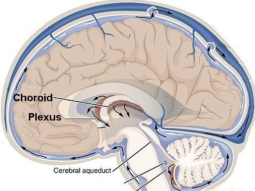
The choroid plexuses are structures of the brain that make the cerebrospinal fluid, the fluid that normally flows around the brain and spine. Sometimes fluid becomes trapped and forms pockets in the choroid plexus. If these pockets are larger than 2 millimeters they are called choroid plexus cysts (CPC).
A choroid plexus cyst (CPC) is not a birth defect, and does not cause any long term health problems or affect development of the baby’s brain .
CPCs may be seen in approximately 1% to 2% of fetuses in the second trimester of
pregnancy .
|

-
https://cnx.org/contents/FPtK1zmh@8.25:fEI3C8Ot@10/Preface
|
Although CPCs may occur alone
(isolated) . when a CPC is found a detailed ultrasound will usually be done on the fetus to
look for a condition called trisomy 18 which has been associated with
the finding of CPCs. Additional findings of a heart defect, clenched
hands, clubbed feet, poor fetal growth , and too much amniotic fluid
suggest the fetus may have trisomy 18 (an extra number 18
chromosome)
If the pregnant person has not already had screening for chromosome abnormalities
(aneuploidies) when a CPC is found, the Society for Maternal-Fetal Medicine (SMFM) recommends counseling to estimate the
chances of trisomy 18 in the fetus and a discussion of screening for
chromosome abnormalities with cfDNA or quad screen
For pregnant people with negative serum or cfDNA screening
results and isolated CPCs, the SMFM recommends no further evaluation
for chromosome abnromalities,"... as this finding is a normal variant of no clinical
importance with no indication for follow-up ultrasound imaging or
postnatal evaluation "
By Mark Curran, MD FACOG Updated 3/5/2022
REFERENCES
1. Peleg D, Yankowitz J. Choroid plexus cysts and aneuploidy. J Med Genet. 1998 Jul;35(7):554-7. doi: 10.1136/jmg.35.7.554. PMID: 9678699
2.
Reddy UM, Abuhamad AZ, Levine D, Saade GR; Fetal Imaging Workshop Invited Participants*. Fetal imaging: executive summary of a joint Eunice Kennedy Shriver National Institute of Child Health and Human Development, Society for Maternal-Fetal Medicine, American Institute of Ultrasound in Medicine, American College of Obstetricians and Gynecologists, American College of Radiology, Society for Pediatric Radiology, and Society of Radiologists in Ultrasound Fetal Imaging workshop. Obstet Gynecol. 2014 May;123(5):1070-1082. doi: 10.1097/AOG.0000000000000245. PMID: 24785860.
3.
Society for Maternal-Fetal Medicine (SMFM). Electronic address: pubs@smfm.org, Prabhu M, Kuller JA, Biggio JR. Society for Maternal-Fetal Medicine Consult Series #57: Evaluation and management of isolated soft ultrasound markers for aneuploidy in the second trimester: (Replaces Consults #10, Single umbilical artery, October 2010; #16, Isolated echogenic bowel diagnosed on second-trimester ultrasound, August 2011; #17, Evaluation and management of isolated renal pelviectasis on second-trimester ultrasound, December 2011; #25, Isolated fetal choroid plexus cysts, April 2013; #27, Isolated echogenic intracardiac focus, August 2013). Am J Obstet Gynecol. 2021 Oct;225(4):B2-B15. doi: 10.1016/j.ajog.2021.06.079. Epub 2021 Jun 23. PMID: 34171388.
|

