|
Reviewed by Medical Advisory Board

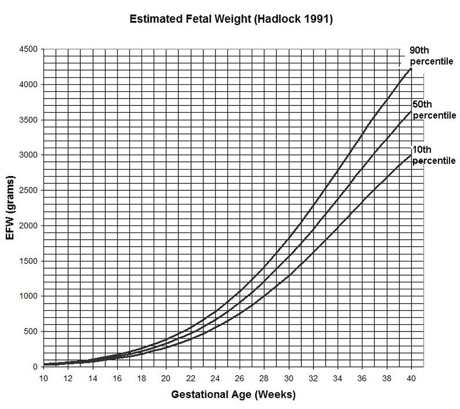
Fetal growth restriction (FGR) , also known as intrauterine growth
restriction (IUGR), is a condition in which an unborn baby (fetus)
has an estimated
fetal weight (EFW) or abdominal circumference (AC) below the 10th
percentile for an accurately assigned gestational age. This means that the baby weighs less
than or has a belly smaller than 9 out of 10 babies of the same gestational age. FGR may be due to
fetal , placental
conditions., or maternal conditions
Causes of Fetal Growth Restriction
Fetal Conditions
Up to 22% of fetuses are naturally small due to the size of the
baby's parents, ethnic background , and the sex of the baby.
About 20% of fetuses have FGR caused by chromosomal or genetic
syndromes, or physical malformations such as heart defects,
diaphragmatic hernia , or gastroschisis .Approximately 5 % of FGR is
caused by congenital infections , with cytomegalovirus (CMV) infection
being the the most common intrauterine infection in the United States.
Multiple gestations account for 3 % of all cases of FGR ; up to 30% of
twins may develop FGR .
Placental
Conditons ( 20 -35%)
Preeclampsia, confined placental
mosaicism, placental mesenchymal dysplasia, placental infarction and decidual vasculopathy, single
umbilical artery, velamentous cord insertion have all been
associated with FGR.
Maternal
Conditons
Maternal consitions that have been associated
with FGR including but not limited to hypertensive disease ,
antiphospholipid syndrome (APLS ),diabetes with vascular
diseases, renal impairment, cigarette smoking,alcohol
consumption, uncontrolled asthma, cystic fibrosis, cyanotic congenital
heart disease, severe anemia, sickle cell anemia, b-thalassemia, and
hemoglobin H disease
CLASSIFICATION OF FGR
- Early onset FGR: FGR diagnosed at less than 32 weeks
- Tends to be more severe and more likely to be assoiated with a
congenital syndrome than late onset FGR.
- Late onset FGR : FGR: idiagnosed at
32 weeks or later
- Accounts for 70% to 80% of FGR cases
and is typically milder than
early onset FGR . Normal Doppler studies of the umbilcal artery
is not uncommon.
- Severe FGR .The EFW is less than 3rd percentile
Classification of FGR as symmetric or
asymmetric based on the head circumference: abdominal circumference
(HC/AC) ratio appears to be of limited value since the HC/AC ratio has
not been found to be an independent predictor of adverse pregnancy
outcomes, or of poor growth or developmental delay in growth restricted
preterm newborns.
EVALUATION
Detailed obstetrical ultrasound to look for malformations
Chromosomal microarray analysis (CMA)* for:
Early-onset FGR
Abnormalies found on ultrasound examinationPolyhydramnios
PCR CMV on amnioc fluid if amniocentesis is done
may require more advanced testing methods for example methylation
analysis, uniparental disomy analysis , deletion / duplication analysis
, sequence analysis
TREATMENT
Currently there are no effective
treatments available for FGR. Activity restriction , and treatment
with heparin or sildenafil are not recommended
MONITORING
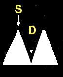
The fetus with FGR is monitored using cardiotocography
(CTG)
and Doppler ultrasound
of the fetal umbilical arteries after viabiity
..
MANAGEMENT
|
FINDINGS
|
Frequency of
UA Doppler |
Frequency
of Cardiotocography |
Frequency
of ultrasound for EFW |
Delivery |
| Normal Doppler |
EFW >= 3rd % or < 10
th% |
Every
week for 2 weeks.
If stable, then every 2 to 4 weeks |
every
week* |
every
3 to 4 weeks |
38 0/7
to
39 0/7 weeks |
| EFW < 3rd % |
every
week |
every
week* |
every
2 weeks |
37 0/7 weeks |
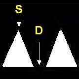
| Decreased EDV |
|
every
week |
1
to 2 times per week* |
every
2 weeks |
37 0/7 weeks |
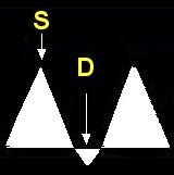
| AEDV |
Consider inpatient admission
Corticosteroids for FLM |
2
to 3 times per week |
2
time per week
if
outpatient* |
every
2 weeks |
33 0/7 to
34 0/7 weeks |
| REDV |
Inpatient admission
Corticosteroids for FLM |
|
1
to 2 times per day* |
every
2 weeks |
30 0/7
to
32
0/7 weeks |
EFW:= Estimated fetal weight; FLM= Fetal lung maturity
Antenatal corticosteroids are indicated if delivery is
anticipated within 7 days in a women at less than 36 6/7 weeks who
has not
received a previous course of antenatal corticosteroids and has no
contraindications . Magnesium sulfate is recommended for
neuroprotection if delivery before 32 weeks is anticipated.
By Mark Curran, MD FACOG Updated
4/12/2021
REFERENCES
1. Giles WB, et al .,Fetal umbilical artery flow velocity waveforms and placental resistance:
pathological correlation. Br J Obstet Gynaecol. 1985 Jan;92(1):31-8.
PMID: 3966988
2. Society for Maternal-Fetal Medicine (SMFM). Electronic address: pubs@smfm.org, Martins JG, Biggio JR, Abuhamad A. Society for Maternal-Fetal Medicine Consult Series #52: Diagnosis and management of fetal growth restriction: (Replaces Clinical Guideline Number 3, April 2012). Am J Obstet Gynecol. 2020 Oct;223(4):B2-B17. doi: 10.1016/j.ajog.2020.05.010. Epub 2020 May 12.
PMID:32407785
3. Longo S, Borghesi A, Tzialla C, Stronati M. IUGR and infections. Early Hum Dev. 2014 Mar;90 Suppl 1:S42-4. doi: 10.1016/S0378-3782(14)70014-3. PMID: 24709457.
4. Lazzarotto T, Guerra B, Gabrielli L, Lanari M, Landini MP. Update on the prevention, diagnosis and management of cytomegalovirus infection during pregnancy. Clin Microbiol Infect. 2011 Sep;17(9):1285-93. doi: 10.1111/j.1469-0691.2011.03564.x. Epub 2011 Jun 1. PMID: 21631642.
5. Medically Indicated Late-Preterm and Early-Term Deliveries: ACOG Committee Opinion, Number 818. Obstet Gynecol. 2021 Feb 1;137(2):e29-e33. doi: 10.1097/AOG.0000000000004245. 33481529
6 Suhag, A., Berghella, V. Intrauterine Growth Restriction (IUGR): Etiology and Diagnosis. Curr Obstet Gynecol Rep 2, 102–111 (2013). https://doi.org/10.1007/s13669-013-0041-z
|