
Placenta previa
Placenta previa is a condition in which the placenta (including the marginal veins of the placenta) partially or completely covers the opening of the cervix (internal cervical os).
|
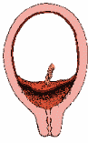
Placenta previa
|
|||
|
|
The placental edge covers the internal cervical os. Follow-up ultrasonography is recommended at 32 weeks of gestation. |
|
|
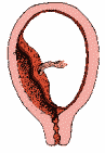
Low-lying placenta
|
|||
|
The placental edge is less than 2 cm from the internal os but does not cover the internal os. Follow-up ultrasonography is recommended at 32 weeks of gestation [4]. |
||
|
LifeART images © 2006 Lippincott Williams & Wilkins.
All rights reserved.
|
|||
In 2014, the Executive Summary of a Joint Eunice Kennedy Shriver National Institute of Child Health and Human Development, Society for Maternal-Fetal Medicine, American Institute of Ultrasound in Medicine, American College of Obstetricians and Gynecologists, American College of Radiology, Society for Pediatric Radiology, and Society of Radiologists in Ultrasound fetal imaging workshop recommended eliminating the terms “partial” and “marginal” and retaining only the terms “placenta previa” and “low-lying placenta” [4].
Placenta previa occurs in approximately 1 in 200 to 250 births overall, but is much more common if a woman has given birth before, has had a cesarean section, has had placenta previa with a previous pregnancy, or is over the age of 35. It is uncommon in nulliparous women (women who have never given birth) [5].
The main symptom of placenta previa is vaginal bleeding. The bleeding is typically painless unless there is coexisting placental abruption or labor. About 20 % of third-trimester bleeding may be attributed to placenta previa [5]. Nearly all cases of placenta previa are delivered by cesarean section. Infrequently, patients with marginal/low-lying placenta and minimal bleeding are allowed to deliver vaginally, as are patients with intrauterine fetal demise (stillbirth) or a previable pregnancy [6].
References