|
Abdomen (The Belly)
The body cavity
below the chest that contains the stomach, liver, intestines and other organs.
Abdominal circumference (AC)
The distance around fetal abdomen.
Read more...
Abortion
Intentional or naturally occurring termination of pregnancy
before the fetus is developed enough to survive.
See related terms
Abruptio placenta (Placental abruption)
Partial or complete separation of the placenta from the uterus before delivery. It
happens in 0.8-1.0% of all pregnancies and has a high recurrence
rate. Contractions are usually present. Bleeding is also present in
approximately 80% of patients.
Factors that have been associated with abruption include maternal hypertension, intrauterine growth restriction (IUGR), non-vertex presentation, polyhydramnios, advanced maternal age, maternal smoking, cocaine use, chorioamnionitis,
premature rupture of membranes, and blunt external maternal trauma
Acceleration
An acceleration is an abrupt increase in the fetal heart rate above baseline
with onset to peak of the acceleration less than 30 seconds and less than 2
minutes in duration.
Acromelia
Shortening of the hands or feet
Active Labor
The active phase (active labor) of labor begins when the cervix is opened
(dilated) to 6 cm in the presence of uterine contractions. During the
active phase uterine contractions become more frequent, the cervix dilates more
quickly, and the baby descends into the pelvis.
Acute Cervical Insufficiency
Cervical dilation of at least 2 cm with membranes visible at 16 0/7 to 22 6/7
weeks' gestation as used by Owen J et al.
Owen J, et al. Multicenter randomized trial of cerclage for preterm birth
prevention in high-risk women with shortened midtrimester cervical length. Am J
Obstet Gynecol. 2009 Oct;201(4):375.e1-8.PMID:19788970
Afterbirth
Collective term for the placenta and fetal
membranes that are delivered after the infant
Agenesis of the corpus callosum (ACC)
A birth defect in which there is partial or complete absence of the corpus
callosum (the bundle of nerve fibers that connects the two hemispheres of the
brain). Read more...
Akinesia
Absence or lack of movement
Alloimmunization (Isoimmunization)
Production of an antibody against antigens produced by members of the same species.
Alpha-fetoprotein (AFP)
A protein produced by the fetal liver and yolk sac that can be detected in
the mother's blood. Alpha-fetoprotein levels rise gradually throughout most of
pregnancy and level off near term. High levels of alpha-fetoprotein are
associated with a more advanced pregnancy than expected, multiple pregnancy,
fetal death (including a vanished twin), an opening in the spine (spina bifida),
an opening in the head (anencephaly), or an opening in the abdominal wall
(gastroschisis). Low levels may be associated with Down syndrome, trisomy
18, and some cases of Turner syndrome.
Amniocentesis
A procedure in which a needle is inserted into the uterus and a sample of the
fluid surrounding the fetus is drawn out. The procedure may be done to evaluate
the fetal chromosomes, to determine fetal lung
maturity, or to obtain fluid to culture for possible infections. The procedure
may also be performed to remove an excessive amount of amniotic fluid.
Amnioinfusion
Infusion of fluid (usually normal saline or lactated Ringer's solution) into the amniotic cavity.
Amniotic fluid
Amniotic Fluid Index (AFI)
Amniotic fluid ‘sludge’
The sonographic finding of dense aggregates of particulate matter in the
amniotic fluid close to the internal cervical os. Amniotic Fluid (AF) ‘sludge’
has been associated with microbial invasion of the amniotic cavity (MIAC), and
histologic chorioamnionitis in patients with spontaneous preterm labor and
intact membranes
Kusanovic JP, et al. Clinical significance
of the presence of amniotic fluid 'sludge' in asymptomatic patients at high risk
for spontaneous preterm delivery. Ultrasound Obstet Gynecol. 2007
Oct;30(5):706-14.
PMID: 17712870
Amniotic sac
The membrane (amnion) that surrounds the fetus and the amniotic
fluid.
Amniotic sheet
A 'shelf' in the amniotic cavity seen during ultrasound examination. Amniotic sheets
represent chorion and amnion that has grown around uterine synechiae (
adhesions) . Incomplete amniotic sheets have a free edge. Complete
amniotic sheets have no free edge, and have been associated with increased risk
for intrauterine death.
Amniotic sheets may be mistaken for amniotic bands. However, amniotic bands more
often appear as multiple thin membranes, and are frequently attached to
the fetus. Circumvallate placenta is another cause of uterine band, sheet, or shelf.
Tan KB, Tan TY, Tan JV, Yan YL, Yeo GS. The amniotic sheet: a truly benign
condition?Ultrasound Obstet Gynecol. 2005 Nov;26(6):639-43.
PMID: 16254890
Amniotomy (artificial rupture of membranes , AROM)
A procedure performed (often using a plastic device that looks like a crochet
needle ) to open the amniotic sac usually for the purpose of inducing or
speeding up the progress of labor .
Anemia
Decreased amount of normal hemoglobin in blood. Hemoglobin is
the substance in red blood cells that carries oxygen.
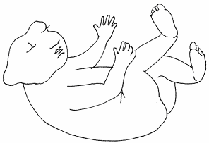
Anencephaly
A birth defect resulting in the absence of a major portion of the skull and
brain. Anencephaly results when the upper portion of the neural tube fails to
close. The condition is not compatible with life, and infants usually die within
a few days after delivery.
Anesthesia
Loss of sensation.
Angioedema, Hereditary
Angle of insonation
A measure of deviation from
"straight on" to a reference plane measured in degrees. For example a Doppler
ultrasound beam aligned to the flow of blood in a vessel has a zero degree of
insonation to the flow. A Doppler
ultrasound beam aligned perpendicular to the flow of blood in the same vessel
has a 90 degree angle of insonation to the flow.
Anomaly
Malformation or abnormality.
Antenatal
Before birth.
Antenatal corticosteroids , ACS
Steroids (either betamethasone or dexamethasone) given to help the fetal lungs and other organs mature more rapidly.
Antenatal steroids are given when preterm delivery is anticipated between 24 and 34 weeks' gestation with intact membranes, and
at 24 to 32 weeks' with ruptured membranes.
Antepartum
Before delivery or birth.
Anterior
In front
Antibody (Immunoglobulin)
Proteins secreted by white blood cells (lymphocytes) that bind to foreign
molecules. Antibodies attach to the antigens and destroy the invader directly ,
or label them for removal by white blood cells in thebody. Antibodies (immunoglobulins) are grouped into five classes or isotypes: IgG,
IgA, IgM, IgD, and IgE.
A molecules that stimulates antibody production is called an antigen ( antibody generator).
Anticardiolipin antibodies (ACA, aCL Antibody) *
An antibody that attaches to cardiolipin , a fatty molecule, found
mostly in the mitochondrial inner membrane where it is synthesized from
phosphatidylglycerol . ACA may be found in several diseases including
antiphospholipid syndrome and systemic lupus erythematosus (SLE). Three classes
of cardiolipin antibodies may be present in the blood: IgG, IgM and/or IgA.
Anti-c antibody (little c antibody) *
A protein made by the immune system that binds to a molecule called the c antigen found on the surface of red blood cells. The c antigen is part of the
Rhesus blood group system which consists of several antigens (D
, E
, e
, c,
C,
). The antibody hastens removal of the c antigen (and the foreign blood cells) from the body.
Anti-c antibody is capable of crossing the placenta and causing anemia in the fetus and
hemolytic disease of
the newborn. Pregnancies complicated by anti-c antibody are managed as for Rh-D sensitization .
Anti-D antibody (Rh sensitization, Rh disease)*
A protein made by the immune system that binds to a molecule
called the D antigen found on the surface of red blood cells. The D antigen is
part of the Rhesus blood group system which consists of several antigens (D
, E
, e
, c,
C,
).
The antibody hastens removal of the D antigen (and the
foreign blood cells) from the body.
Anti-D antibody is capable of crossing the placenta and causing SEVERE anemia in the fetus and
hemolytic disease of
the newborn.
Anti-Duffy antibody (anti-Fya antibody)
A protein made by the immune system that binds to a molecule called the Fya
antigen found on the surface of red blood cells. The Fya antigen is part of the Duffy blood group system
which consists of the antigens Fya and
Fyb . The antibody hastens removal of the and Fya antigen (and the foreign blood cells) from the body.
Anti-Fya antibody is capable of crossing the placenta and causing SEVERE anemia in the fetus and
hemolytic disease of
the newborn. Anti-Fyb has not been reported to cause significant hemolytic disease of
the newborn.
Anti-Kell antibody
A protein made by the immune system that binds to a molecule called the Kell antigen found on red blood cells. The Kell
antigen is part of the Kell blood group system which consists of several antigens ( Kell
or K1 , Kpa,
k
, Jsa
,Jsb ). The antibody hastens removal of the Kell antigen (and the foreign blood cells) from the body.
Anti-Kell antibody is capable of crossing the placenta and causing SEVERE anemia in the fetus and
hemolytic disease of the newborn.
SEE ALSO: Q & A ..a pregnant patient with
anti-Kell antibodies...
Anti-Kidd antibody (anti-Jka or anti-Jkb)
A protein made by the immune system that binds to a molecule called Kidd
antigen found on the surface of red blood cells.
The Kidd antigens Jka
and Jkb
are part of the Kidd blood group system.
Anti-Kidd antibody is capable of crossing the placenta and causing anemia in the fetus and hemolytic disease of
the newborn.
Anti-Lewis antibody
A protein made by the immune system that binds to molecules called the Lewis
antigens,
Le
a and Le b. Lewis antigens are not made by the red blood
cell, but are antigens present in body fluids and secretions that have been
adsorbed onto the surface of the red blood cell.
Lewis antigens are found in very low levels on the fetal red cells.
Most Lewis antibodies are of the IgM type and do not cross the placenta.
Lewis blood group antibodies are not known to cause
hemolytic disease of
the newborn.
Anti-S antibody
A protein made by the immune system that bind to a molecule called the S antigen found on the surface of red blood cells.
The S antigen is part of the MNS blood group system which consists of several antigens (
M,
S,s,
N)
Anti-S antibody is capable of crossing the placenta and causing anemia in the fetus and
hemolytic disease of
the newborn.
Apgar Score
Apnea
Temporary cessation of breathing
Arcuate uterus
Midline
thickening of the wall of the uterus at the uterine fundus (top of the
uterus). The thickened area results from failure to completely dissolve the
uterine septum during development. The arcuate uterus is considered to be a mild form of
bicornuate uterus.
An arcuate uterus does not appear to have an unfavorable effect on
pregnancy.
|
 |
Areola
The darker colored area around the nipple of the breast
Arnold-Chiari Malformation
A group of birth defects of the cerebellum (the part of the brain that
controls balance) and base of the skull characterized by downward displacement of the cerebellum and
related structures below the level of the foramen magnum (the large hole at the
base of the skull).
Arrest of descent
Second-stage arrest may be diagnosed if there has been
"No progress (descent or rotation) for
4 hours or more in nulliparous women with an epidural
3 hours or more in nulliparous women without an epidural
3 hours or more in multiparous women with epidural
2 hours or more in multiparous women without an epidural"
Spong CY, et. al. Preventing the first cesarean delivery: summary of a joint
Eunice Kennedy Shriver National Institute of Child Health and Human Development,
Society for Maternal-Fetal Medicine, and American College of Obstetricians and
Gynecologists Workshop. Obstet Gynecol. 2012 Nov;120(5):1181-93. doi
http://10.1097/AOG.0b013e3182704880. PMID: 23090537
Arrest of dilatation
For spontaneous labor:
6 cm or greater dilation with membrane rupture AND
4 hours or more of adequate contractions (e.g., > 200 Montevideo units) OR
6 hours or more if contractions inadequate with no cervical change
For induced labor:
6 cm or greater dilation with membrane rupture or 5 cm or greater without
membrane rupture AND
4 hours or more of adequate contractions (e.g., > 200 Montevideo units) OR
6 hours or more if contractions inadequate with no cervical change [4].
Spong CY, et. al. Preventing the first cesarean delivery: summary of a joint
Eunice Kennedy Shriver National Institute of Child Health and Human Development,
Society for Maternal-Fetal Medicine, and American College of Obstetricians and
Gynecologists Workshop. Obstet Gynecol. 2012 Nov;120(5):1181-93. doi
http://10.1097/AOG.0b013e3182704880. PMID: 23090537
Artificial rupture of membranes (AROM)
See amniotomy.
Arthrogryposis
Ascites
Aspirate To inhale into liquid or food into the lungs, or to draw
(fluid) by suction
Augmentation of
labor
Stimulation the uterus to increase the frequency, duration , or strength of
contractions when spontaneous contractions have failed to cause dilation or thinning (effacement) of the cervix leading to the delivery of the infant.
1. World Health Organization. Managing Complications in Pregnancy and Childbirth .A guide for midwives and doctors. 2003. http://who.int/reproductive-health/impac/Procedures/Induction_P17_P25.html
ACOG Practice Bulletin No. 49.
2. American College of Obstetricians and Gynecologists. Dystocia and augmentation of labor.Obstet Gynecol 2003;102:1445–54.
Autologous transfusion
"Infusion of blood or blood component to the same individual from whom it
was taken"
Autosomal dominant
A trait determined by a gene on any chromosome other than a sex chromosome
(X or Y) that requires only one gene for the trait to be expressed. The chance
of passing the trait to an offspring is at least 50% for each pregnancy.
See Diagram
Autosomal recessive
A trait determined by a gene on any chromosome other than a sex chromosome
(X or Y) that requires two genes for the trait to be expressed. A person with
only one copy of the gene is said to be a carrier for the trait.
See Diagram
Bag of waters
The membrane (amnion) surrounding the fetus and the amniotic fluid.

Bicornuate Uterus
Two separate single horn uterine bodies sharing one cervix.
Bicornuate uterus is associated with increased risk for miscarriage, preterm
labor, breech presentation, and fetal growth restriction.
Biophysical profile (BPP)
Bishop Score
The Bishop Score (also known as Pelvic Score)
method used to rate
the likelihood of a woman entering labor naturally in the near future.
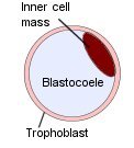
Blastocyst
The developmental stage of the fertilized egg about 5 days after
fertilization before it implants into the lining of the uterus. It consists of
a a liquid filled sphere with an outer layer of cells, the trophoblast, that
will form the placenta and an inner cell mass that forms the embryo.
Blighted Ovum
A fertilized egg that has failed to develop.
Blood
pressure
Bloody show
Passage of blood-tinged mucus from the vagina caused by loss of the
cervical mucous plug. Bloody show often precedes the onset of labor.
Body Mass Index (BMI)
The body mass index (BMI) is a measure of someone's weight in relation to
their height. The BMI is used to estimate the amount of body fat a person has.
The BMI is calculated either as:
BMI = (Weight in pounds / Height in inches 2 ) x 703
OR
BMI = (Weight in kilograms / Height in meters 2)
Bradley Method (husband-coached birth)
A method of natural childbirth developed by Robert A. Bradley, M.D.
(1917–98). The Bradley method emphasizes education and relaxation techniques
for pain management. The method prepares the baby's father to be the mother's
birth coach, and prepares the mother to deliver without pain medication.
Bradycardia
In a fetus a mean heart rate less than 110 beats per minute lasting for at
least two minutes. In an adult a sustained heart rate less than 60 beats per
minute.
Braxton Hicks Contractions
Sporadic uterine contraction that do not increase in intensity and do not
result in childbirth, typically felt after 20 weeks. Named after John Braxton
Hicks a British gynecologist who first described these contractions in 1872.
Breech presentation
The baby is in a sitting position with the buttocks, knees, or feet nearest to
the cervix.
Breech presentation occurs in 25 percent of pregnancies less than 28 weeks'
and 1 to 3 percent of births at term. The three types of breech presentation
are frank breech (flexed at hips with extended knees-legs above buttocks),
footling breech (one or both hips extended-leg(s) extended below buttocks),
and complete breech (flexed hips and knees-no limbs extended).
Campomelia
Bowing of the long bones
Caput succedaneum
Swelling and accumulation of fluid (edema) in the scalp of infants born
vaginally. The swelling usually disappears within 24 to 48 hours.
Carpal Tunnel Syndrome (CTS)
Pain, numbness , and weakness of the hand and fingers caused by pinching of
the median nerve as it passes through the space over the wrist (carpal) bones
. Conditions associated with carpal tunnel syndrome include diabetes,
hypothyroidism, arthritis, obesity, and pregnancy.
Catheter
A hollow tube used to inject fluid into, or drain fluid from a space such as
the bladder.
Cephalhematoma
A collection of blood caused by rupture of blood vessels between the skull and
the periosteum (the membrane surrounding a bone). The blood does not cross the
joints of the skull, because it is trapped between the periosteum and bone.
Subtle skull fractures may underlie a cephalhematoma. The condition generally
resolves over several weeks.
Cephalic presentation
The fetal head is down near the mother's cervix.
Cephalic index
The ratio of the bi-parietal diameter (BPD) to the occipito-frontal diameter
(OFD) X 100. The normal range is 70 to 86. A cephalic index of less than
70 is
considered dolichocephaly. An index of greater than 86 is considered to be
brachycephalic
Cephalhematoma
A collection of blood caused by rupture of blood vessels between the skull and
the periosteum (the membrane surrounding a bone). The blood does not cross the
joints of the skull, because it is trapped between the periosteum and bone.
Subtle skull fractures may underlie a cephalhematoma. The condition generally
resolves over several weeks.
Cephalopelvic disproportion (CPD)
The fetal head is too large to pass through the mother's pelvis. Cephalopelvic disproportion is usually diagnosed
when labor fails to progress (cervical dilation and effacement have stopped) and
is unresponsive to oxytocin
augmentation.
Cerclage
A procedure used to
temporarily stitch the cervix closed in pregnant women with a history of
premature delivery caused by an incompetent cervix. Cerclage
sutures are usually placed at 10 to 15 weeks' gestation
Cerebral Palsy
A group of disorders characterized by inability to move and /or to control
movements caused by injury or abnormal development in the immature brain. Affected individuals may have loss or impairment of normal movement, spasms, difficulty swallowing or speaking, impaired vision or hearing, and seizures
Certified nurse midwife (CNM)
A registered nurse with at least 1-2 years of nursing experience who has
received additional training in delivering babies and providing prenatal and
postpartum care to women. They are certified by the American College of
Nurse-Midwives (ACNM).
Cervical os
The opening of the cervix
Cervical incompetence
Cervical insufficiency (sometimes called an incompetent cervix) is the failure
of the cervix to maintain a pregnancy when there are no signs or symptoms of
labor in the second trimester.
Cervical ripening
The process where the cervix becomes ready for labor by becoming softer ,
thinner and opening (dilating) during the last few weeks of pregnancy.
Medications or mechanical dilators are sometimes used to artificially ripen
the cervix before induction to make the cervix more favorable and a vaginal
delivery more likely
Cervix
Lower narrow part of the uterus that opens into the vagina.
Cesarean section (C-section)
An incision made through the abdomen and uterus for the purpose of delivering
one or more fetuses. The incision on the abdomen may be vertical or
transverse. The incision made on the uterus may not be in the same direction
as the abdominal incision.
- Low
transverse (Kerr) See illustration
- The
most common incision. This incision is easy to repair and is associated with
the lowest probability of rupture or dehiscence in a subsequent pregnancy
- Low
vertical (Kronig) See illustration
- Used
when lower uterine segment is undeveloped or for premature breech
presentation.
-
Classical See illustration
- This incision may be used when a back down transverse lie
that cannot be converted to breech or cephalic presentation, inability to
expose the lower uterine segment, premature breech presentation, and
anterior placenta previa.
Chadwick's sign
Bluish discoloration of the vaginal tissue and cervix caused by
accumulation of blood (venous congestion). Chadwick's sign may be seen as
early as six weeks of pregnancy.
CHARGE syndrome
A congenital disorder characterized by
Coloboma,
Heart defects,
choanal
Atresia,
Retarded growth and development,
Genital abnormalities, and
Ear anomalies.
Lalani SR, Hefner MA, Belmont JW, et al. CHARGE Syndrome. 2006 Oct 2
[Updated 2012 Feb 2]. In: Pagon RA, Adam MP, Ardinger HH, et al., editors.
GeneReviews® [Internet]. Seattle (WA): University of Washington, Seattle;
1993-2016. Available from:
http://www.ncbi.nlm.nih.gov/books/NBK1117/
Chemical pregnancy
A positive pregnancy test ( elevated hCG level in the blood or urine) before
pregnancy can be verified by ultrasound. Often used to refer to a pregnancy that
has failed before reaching a size large enough to be seen on sonogram.
Chloasma (mask of pregnancy, melasma)
Blotchy areas of darkened skin over the the forehead, cheeks and upper lips
associated with pregnancy or with the use of contraceptives. Exposure to
ultraviolet (UV) rays from the sun or tanning salons intensifies the pigment
changes. The areas of darkened skin usually fade several months after delivery
or discontinuation of the contraceptive
Cholestasis of pregnancy (Intrahepatic cholestasis of pregnancy ,ICP)
A liver disorder in which the release of bile from the liver is thought to be
blocked by high levels of the hormones estrogen and progesterone during
pregnancy. The bile builds up in the blood causing itching. Cholestasis of
pregnancy usually occurs in the second half of pregnancy, and affects about
one in 200 pregnancies.
Chorioamnionitis
Inflammation of the fetal membranes and amniotic fluid usually associated with
a bacterial infection. The bacteria responsible are usually those that are
normally present in the vagina. The presence of fever, uterine tenderness, and
foul vaginal discharge help to confirm the clinical diagnosis of
chorioamniotis.
Chorion
The outermost of the two fetal membranes that gives rise to the placenta.
Chorionic Villus Sampling (CVS)
Removal of cells that line the placenta, the chorionic villi, through the
cervix using a catheter or through the abdomen using a needle. The material
obtained may be tested for Down syndrome and other disorders. The procedure is
usually performed between the 10th and 12th weeks of pregnancy .
Choroid plexus
Structures in the ventricles (spaces) of the brain that produce the
cerebrospinal fluid. Each plexus is made up of a network of capillary blood
vessels covered by transporting epithelial cells.
Choroid plexus cyst
Pockets of fluid in the choroid plexus believed to be caused by abnormal
folding of the epithelium lining of the choroid plexus which traps fluid and
debris .
Chromosome
Structures made of of tightly coiled DNA (deoxyribonucleic acid) found in the
nucleus of a cell.
Chromosomes are the structures in the cells in the body that are inherited
from each of parent, and hold the instructions for the body looks
and functions. Humans have 23 pairs of chromosomes for a total of 46. The
first 22 chromosomes are numbered from largest to smallest in size. The 23rd
pair are the sex chromosomes and are named as X or Y .
Chronic hypertension
High blood pressure
present before
pregnancy or detected before 20th week of pregnancy.

Circumvallate placenta
The membranes insert closer to the center of the placenta instead of extending
to the edge of the placenta creating a folded and thickened placental margin that appears as a 'shelf-like'
structure at the placental edge during ultrasound examination. Circumvallate placenta has been associated with premature labor, stillbirth,
hemorrhage and placental abruption
1.
Suzuki S.Clinical significance of pregnancies with circumvallate placenta.J
Obstet Gynaecol Res. 2008 Feb;34(1):51-4.
PMID: 18226129
2.
Harris RD, et al. Accuracy of prenatal sonography for detecting circumvallate
placenta. AJR Am J Roentgenol. 1997 Jun;168(6):1603-8.
PMID: 9168736
Cleft lip and palate (orofacial cleft)
A gap of the lip or lip and palate (roof of the mouth) caused by failure of
the lip or the lip and palate to grow together.
The lip and primary palate
close during the 4th to 7th weeks of gestation.
The secondary palate begins to close the 6th week and is completed
between the 9th and 12th weeks of gestation. Cleft lips are unilateral
or bilateral. See
Image
Club foot (Talipes equinovarus)
The foot is turned inward. Both feet are affected in 50% of cases. The defect
may be corrected surgically. Club foot occurs in about 1 in 700 to 800 births.
In a small number of cases, clubfoot may be seen in association with spina
bifida or as part of a skeletal dysplasia. The estimated risk of recurrence in
future children is 3 to 8% if 1 child is affected and 10% if 1 child and 1
parent are affected.
Colostrum
Thin, yellow, milky fluid secreted by the breasts in the last weeks of
pregnancy and the first few days after delivery. Colostrum contains high
levels of maternal antibodies.
Colpocephaly
Enlargement of the occipital horns of the lateral ventricles in the brain.
Congenital Pulmonary Airway Malformation (CPAM ) , Congenital Cystic
Adenomatoid Malformation (CCAM)
An abnormally formed piece of lung made up of closed sacs (cysts) that will
never function as normal lung tissue . A CPAM usually involves only one lobe
of a lung. We do not know what causes CPAM, but CPAM occurs in about 1 in
10,000 fetuses. CPAM was previously called congenital cystic adenomatoid
malformation (CCAM).
On ultrasound examination a CPAM may appear macrocystic, microcystic, or a
mixture of the two. A macrocystic CPAM has one or more large cysts (>= 5mm)
that appear as empty spaces or holes in the lung . A microcystic CPAM has very
small cysts (< 5mm ) giving it the appearance of a solid well-defined mass
that is brighter (more white) than the surrounding normal lung.
Consanguinity
To be related through a recent common ancestor ( a close blood relative ).
Contraction, uterine
Tightening of the muscular wall of the uterus that may feel like menstrual
cramps.
Cord compression, Umbilical cord compression
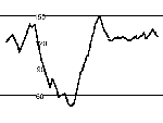
Squeezing or pinching of the umbilical cord . Umbilical cord compression may
interrupt blood flow to the fetus causing the fetal heart rate to decrease .
Decreased heart rate for a brief time
(variable deceleration) may also be seen with
head compression and with movement in the premature fetus.
Sorokin Y, et. al., The association between fetal heart rate
patterns and fetal movements in pregnancies between 20 and 30 weeks' gestation.
Am J Obstet Gynecol. 1982 Jun 1;143(3):243-9. PMID: 7081342
Corpus luteum (CL)
A
yellow colored structure that the develops from cells of the empty egg
follicle after the egg is released.
The corpus luteum secretes progesterone which prepares the lining of the
uterus for implantation by the embryo.
Craniosynostosis
Premature closing of joints or sutures in the skull. Craniosynostosis may
occur as an isolated finding or may be associated with a syndrome
such as Apert, Chotzen, Pfeiffer, Carpenter, and Crouzon syndromes
Crowning
The appearance of the infant's scalp at the vaginal opening during labor.
Crown-rump length (CRL)
The distance between the top of the head (crown) and buttocks (rump) of the
embryo or fetus.
Cystic fibrosis
A condition characterized by thick mucus build up in the lungs and digestive
tract. The mucus in the lungs causes inflammation and infections leading to
the formation of scar tissue (fibrosis) and cysts in the lungs. Cystic
fibrosis (CF) also affects the pancreas, liver, intestines, sinuses, and sex
organs
Cystic hygroma
Single or multiple sac-like structures caused by abnormal development of the
lymphatic system (the system responsible for carrying white blood cells that
help fight infection and disease). Cystic hygromas occur most often about the
neck. More than half of fetuses with cystic hygromas diagnosed in utero have
Turner syndrome (one x chromosome).
Image 1
Image 2
Cytomegalovirus (CMV) infection
Cytomegalovirus (CMV) is a common virus transmitted by direct person-to-person
contact through saliva, breast milk, or urine. About 33% of (33 of every 100)
women who become infected with CMV for the first time during pregnancy pass
the virus to their fetuses. Severe infections can lead to significant damage
to the nervous system and other vital organs of the unborn baby.
Findings on ultrasound that would raise the possibility of a severe CMV
infection include very high or very low levels of amniotic fluid , fluid
collections in the abdomen (ascites), dense appearing (echogenic) bowel,
growth restriction, very small head (microcephaly), dilation of the fluid
filled chambers of the brain ventriculomegaly or hydrocephaly), or calcium
deposits in the brain or liver.
Deep vein thrombosis, DVT
A blood clot in a blood vessel that carries blood back to the heart (vein).
Symptoms include pain, tenderness, and swelling of the affected extremity.
Diabetes
A condition in which a person has an abnormally high amount of sugar (glucose)
in their blood. Diabetes occurs when the body does not produce insulin, the
substance in the body that lowers blood sugar, or the cells in the body do not
respond to insulin .
Diabetic Ketoacidosis(DKA)
A condition that occurs in diabetics due to lack of insulin. Signs and
symptoms may include nausea, vomiting, abdominal pain, thirst, excessive urine
output, dehydration fast pulse, and low blood pressure . Without
treatment DKA progresses to coma and death.
Read more...
Diamniotic
Two separate amniotic sacs (bags of water)
Diaphragmatic hernia (congenital diaphragmatic hernia -CDH)
An abnormal opening in the diaphragm (the muscle used for breathing . It
divides the chest from the abdomen.) caused by failure to completely form the
diaphragm. The defect allows the abdominal organs to move into the chest
cavity which may prevent normal development of the lungs. The condition is
associated with a 30 to 60% death rate due to underdeveloped lungs and
associated abnormalities such as heart defects, malformed or absent kidneys,
and hydrocephalus. The presence of the liver in the chest generally increases
the likelihood of a poor outcome.
Dichorionic
Two separate placentas.
Dilation and curettage (D and C)
A surgical procedure in which the cervix is gradually opened with instruments
called dilators and the surface of the endometrium (lining of the uterus) is
scraped away with a curette, a sharp-edged instrument.
Down syndrome (trisomy 21)

A disorder characterized by mental retardation, flat facial profile with
protruding tongue, poor muscle tone, excess skin on neck, slanting eye
openings (slanted palpebral fissures), abnormal pelvis, and short stature. In
addition there may be heart defects (AV canal defect) , gastrointestinal
malformations, problems with vision and hearing, and increased susceptibility
to leukemia and infections. The syndrome is named after John Langdon Down, the
first physician to identify the syndrome.
Down syndrome occurs in one out of 800 live births and is caused by extra
material from chromosome 21. In most cases (95%) there are three copies of
chromosome 21 instead of two. In 90% of these cases the extra chromosome is
inherited from the mother.
Due date (estimated due date-EDD)
The date that spontaneous onset of labor is expected to occur. The due date
may be estimated by adding 280 days to the first day of the last
menstrual period (LMP).
Dystocia
Slow or difficult labor caused by inadequate uterine contractions,
abnormalities in the maternal pelvis, a large fetus or a combination of these
causes.
Doppler ultrasound
A method using ultrasound to detect and measure blood flow.
Echogenic (hyperechogenic) bowel
Intestine that reflects more sound on an ultrasound examination than usual
making it appear very white. The finding of echogenic bowel may be a normal
variant in some babies. However, the finding of echogenic bowel has been associated with an
increased risk for chromosomal abnormality (such as Down syndrome) , cystic
fibrosis, viral infection (CMV and parvovirus) , unexplained fetal death,
growth restriction, and premature birth.
Echogenic focus
A distinct area that reflects more sound on an ultrasound examination than
usual making it appear very white. The term commonly refers to bright spots
seen in the ventricles of the heart. Very bright small spots may represent
dense papillary muscles or tendons within the heart. Cardiac tumors may also
appear as spots within the heart . However, tumors tend to be larger,
multiple, and are not as bright as an echogenic focus.
Eclampsia
New-onset convulsions (grand mal seizure) in a woman with preeclampsia.
Preeclampsia is a condition characterized by high blood pressure and protein
in the urine that develops after the 20th week of pregnancy. The cause of
preeclampsia is unknown.
Ectopic pregnancy
A pregnancy growing outside of the uterus.
Edema
Swelling caused by the accumulation of fluid under the skin.
Edwards' syndrome (Trisomy 18)
A rare disorder that happens when the baby has three copies of chromosome 18
instead of the usual 2 copies. Chromosomes are the structures in the cells of
your body that are inherited from each of your parents. Babies with
trisomy 18 have severe mental retardation and usually have many birth defects,
because of the extra chromosome 18. Only 5% to 10 % of infants survive the
first year after delivery. Death is usually caused by inability to maintain
normal breathing or heart and lung problems Ultrasound findings that are often
seen in babies with trisomy 18 include cleft lip and palate, a small jaw, low
set ears, club feet, clenched fists, a single umbilical artery , kidney
abnormalities, poor growth, and a high level of amniotic fluid
(polyhydramnios) . More than 90% of babies with trisomy 18 will have a heart
defect.
Effacement
Thinning or shortening of the cervix
Embryo
A fertilized egg from initial cell division until the eighth week of
development.
Encephalocele
A defect affecting the skull resulting in the herniation of the meninges
and portions of the brain through a bony midline defect in the skull
Epidural
A method of pain relief in which anesthesia is injected into the space around
the spinal cord (epidural space)
Episiotomy
An incision made between the vagina and rectum to widen the vaginal opening
for delivery.
Erythema infectiosum (Parvovirus infection)
Erythema infectiosum also known as Fifth disease is a common childhood illness
caused by a virus called parvovirus B19. About 50% of all adults have been
infected sometime during childhood or adolescence. Women who become
infected with parvovirus for the first time during their pregnancy may pass
the virus to their unborn child. Parvovirus can cause severe anemia in the
fetus which may lead to congestive heart failure. The heart itself may become
enlarged. In addition parvovirus infection has uncommonly been associated with
enlarged ventricles in the fetal brain and calcium deposits in the spleen.
External cephalic version
To manually turn the fetus from a breech (sitting position) presentation to a
cephalic presentation (head down nearest to the cervix) by applying external
pressure on the mother's abdomen.
Extremely low birth weight (ELBW)
A birth weight of less than 1000 grams ( 2 pounds 3 ounces)
Factor V
Factor V is a protein in the blood that promotes clotting of the blood by accelerating the activation of prothrombin to thrombin. Activated factor V is inactivated by activated protein C. Factor V is broken down by activated protein C (APC) which acts
to control the formation of the clots.
Factor V Leiden is a form of factor V that is resistant to APC . People
with factor V Leiden have an increased tendency to form blood clots (thrombophilia)
Factor V Leiden Mutation (activated protein C resistance)
A genetic mutation in the factor V gene that makes the activated factor V
protein resistant to inactivation by protein C. The increased activity of
factor V in the blood leads to a higher risk of forming a blood clot (thrombophilia)
. The factor V Leiden mutation has a prevalence of 5–9% in the general
population.
Failed Induction of Labor
"Failure to generate regular (e.g. every 3 minutes) contractions and
cervical change after at least 24 hours of oxytocin administration, with
artificial membrane rupture if feasible. "
Spong CY, et. al., Preventing the
first cesarean delivery: summary of a joint Eunice Kennedy Shriver National
Institute of Child Health and Human Development, Society for Maternal-Fetal
Medicine, and American College of Obstetricians and Gynecologists Workshop.
Obstet Gynecol. 2012 Nov;120(5):1181-93. doi:
http://10.1097/AOG.0b013e3182704880. PMID: 23090537
Failure to progress, prolonged labor
SEE LABOR
Fetal fibronectin (fFN)
Fetal fibronectin (fFN) is a substance that acts like "glue" holding the fetal
sac to the uterine lining during pregnancy. It can normally be found in the
cervicovaginal secretions of women up to 22 weeks of gestation. However,
the presence of fetal fibronectin in cervicovaginal secretions between 24 and
34 completed weeks of gestation is reported to be associated with preterm
delivery.
Fetus
A human conceptus from 70 days' gestational age until delivery
Fetal viability
The capacity for sustained survival outside the uterus as determined by the
judgment of the responsible attending physician. Newborns with
malformations incompatible with life such as renal agenesis, anencephaly,
trisomy 13 , or trisomy 18 are not considered viable regardless of the
gestational age.
Fetal Warfarin Syndrome
A group of birth defects associated with exposure of the developing fetus to
warfarin (Coumadin®) an oral anticoagulant used to prevent blood clots. The
defects include nasal hypoplasia, stippled epiphyses, limb deformities, mental
retardation, and seizures. Hall et al. placed the critical period for
warfarin effects on the fetus as 6 to 9 weeks following conception.
Hall JG , et al. Maternal and fetal sequelae of anticoagulation during pregnancy.Am J Med. 1980 ;68:122-40. PMID: 6985765
Fibroid (leiomyoma, myoma)
A non-cancerous (benign) growth of fibrous tissue and muscle fibers. Fibroids
are often found in the uterus, and occur in about 30% of women over the age of
30.
Fluorescence in situ hybridization, FISH
A technique used for identifying a specific DNA sequence on a chromosome. The
technique uses single-stranded DNA segments attached to a fluorescent
(glowing) dye, called a probe. The probe is mixed with the test chromosomes
and sticks to its corresponding sequence (the DNA sequence being tested for)
on the chromosome.
Follicle stimulating hormone (FSH)
A hormone (a chemical) produced by the pituitary gland in the brain that stimulates the growth of eggs in the ovaries and the production of sperm in the testes. An elevated FSH suggests partial or complete ovarian failure.
Fontanel ("soft spot")
A membranous gap between the bones of the developing skull.
The newborn normally has six fontanels the anterior, posterior, two mastoid,
and two sphenoid fontanels. The most prominent fontanel is the diamond-shaped
anterior fontanel which is closed in 96 percent of infants by 24 months of
age.
Fragile X Syndrome (Martin-Bell syndrome)
A disorder affecting males and females characterized by mental retardation..
In the U.S. fragile X syndrome affects approximately 1 in 4000 males and 1 in
8000 females and is a common cause of autistic-like behaviors. Fragile X
syndrome is caused by an expansion mutation that prevents the normal
expression of the the Fragile X mental retardation 1 (FMR1) gene located on
the X chromosome.
Fraternal twins (dizygotic twins)
Two offspring created when two separate eggs are fertilized by separate sperm
during the same menstrual cycle. The two products of conception develop as two
separate embryos, and each embryo develops its own membranes and placenta.
Fundal height
The distance from the top of the pubic bone to the top of the pregnant uterus
(fundus) in centimeters. The measurement is used to approximate fetal age and
growth.
Fundus
The uppermost part of the uterus.
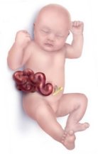
Gastroschisis
Gastroschisis is a birth defect in which the baby's intestines (bowels) stick
out through a hole to one side of the belly button.
Gastroschisis is thought to arise from disruption of blood flow to the
affected abdominal wall. Another explanation is that the yolk sac does
not become part of the the umbilical cord, as it normally does, leaving a hole
about 2 to 4 centimeters (0.8 to 1.6 inches) wide that is nearly always to the
right side of the belly button.
Gene
A section of the molecule DNA (deoxyribonucleic acid) which codes for a
particular protein and carries the hereditary information for such
characteristics as hair color, eye color, and susceptibility to disease.
Genetic counseling
Evaluation of prospective parents' risks of having a child born with a birth
defect or genetic disorder, and advise on their options for testing and
treatment.
Gestational age
The time elapsed since the first day of the last menstrual period. If pregnancy
was achieved using assisted reproductive technology, gestational age is
calculated by adding 2 weeks to the conceptional age.
Gestational diabetes (GDM)
Gestational diabetes mellitus (GDM) is diabetes that is found for the first
time when a woman is pregnant. The high blood sugar in gestational diabetes
appears to be caused by hormones produced by the placenta that prevent the
mother's cells from responding to her insulin. It is estimated that
gestational diabetes affects about 18% of pregnancies.
Gestational hypertension
High blood pressure occurring for the first
time after 20 weeks gestational age
A systolic blood pressure (SBP) greater than or equal to 140 mm Hg OR
a diastolic blood pressure (DBP) greater than or equal to 90 mm Hg on at
least two occasions at least 4 hours apart
The American College of Obstetricians and Gynecologists. Task Force on
Hypertension in Pregnancy Hypertension in Pregnancy. Hypertension, Pregnancy
Induced --Practice Guideline wq244 2013
http://www.acog.org/~/media/Task%20Force%20and%20Work%20Group%20Reports/
Gravida
A pregnant woman.
Group B streptococcus:
Group B streptococcus (GBS) is a bacteria normally found in the vagina and/or
rectum of about 1 in 4 of all healthy women. GBS bacteria passed from the
mother to the baby can cause some babies to become very sick and even die.
Hegar's sign
Softening of the lower uterine segment just above the cervix seen as a probable sign of pregnancy. Originally described by the German gynecologist Ernst Ludwig Alfred Hegar; Hegar's sign may be observed as early as six weeks of pregnancy.
HELLP syndrome
HELLP is an acronym that describes the syndrome of : H Hemolysis;
EL elevated liver enzymes; LP, low platelets.
HELLP syndrome usually presents in the third trimester with right upper
quadrant or epigastric pain, nausea, and vomiting. HELLP syndrome is
considered to be a variant of preeclampsia. HELLP syndrome occurs in
approximately 0.2 to 0.6 percent of all pregnancies. The cause of HELLP
syndrome is unknown.
Strict criteria for the diagnosis of HELLP syndrome:
- Hemolysis (characteristic peripheral blood smear) and serum lactate
dehydrogenase levels >600 U/L
- Serum aspartate aminotransferase levels >70 U/L
- Platelet count <100,000/mul.
Hemolytic disease of the newborn (HDN)
Anemia in a newborn infant caused by the destruction of red blood cells. In
severe cases jaundice, pallor, an enlarged spleen, or hydrops may be present
Hemophilia
A group of hereditary disorders characterized by prolonged bleeding and
sometimes excessive bleeding. Read
more...
Hepatitis B
Hepatitis C
Hepatitis C virus (HCV) is a single-stranded RNA virus . Injecting-drug use currently accounts for 60% of HCV transmission in the United States. Blood transfusion, is an uncommon cause of recently acquired infections . Sexual transmission of HCV appears to be inefficient relative to hepatitis B virus (HBV). Transmission between sexual partners of persons with chronic HCV infection with no other risk factors for infection is about 5% (range, 0% to 15%)
. Approximately 7-8% of hepatitis C virus-positive women transmit hepatitis C virus to their offspring with a higher rate of transmission seen in women coinfected with HIV . In one small study acute maternal hepatitis appears to have no effect on the incidence of congenital malformations, stillbirths, abortions, or intrauterine malnutrition. However, acute hepatitis may increase the incidence of prematurity. Pregnancy does not appear to be adversely affected by chronic HCV.
Hydrocephaly (hydrocephalus, water on the brain)
Enlargement of the spaces within the brain (ventricles ) caused by excessive
fluid (cerebrospinal fluid). The excessive fluid may cause enlargement of the
infant's head.
The abnormally increased fluid may be the result of increased production of
fluid, but more commonly is caused by obstruction of fluid flow between the
different spaces in the brain. Hydrocephaly has been associated with
aqueductal stenosis, spina bifida, X-linked hydrocephalus, Arnold-Chiari
malformation , Dandy-Walker malformation,agesnesis of the corpus callosum , tumors, subarachnoid hemorrhage,
infections (CMV and toxoplasmosis) , and chromosome abnormalities
Hydronephrosis
Enlargement of the renal pelvis (the part of the kidney that collects urine)
to greater than 10 mm. Renal pelvis dilation of 4 to 10 mm in
anterioposterior diameter is commonly referred to as fetal pyelectasis.
Hydronephrosis is usually caused by a blockage of the flow of urine along the
urinary tract.
Hydrops
Hydrops fetalis is a condition in the fetus characterized by an abnormal
collection of fluid with at least two of the following:
- Edema (fluid beneath the skin, more than 5 mm).
- Ascites (fluid in abdomen)
- Pleural effusion (fluid in the pleural cavity, the fluid-filled space
that surrounds the lungs)
- Pericardial effusion (fluid in the pericardial sac, covering that
surrounds the heart)
Read more...
Hypertension
High blood pressure . A systolic blood pressure (SBP) greater than or equal
to 140 mm Hg OR a diastolic blood pressure (DBP) greater than or equal to 90 mm Hg or both
The American College of Obstetricians and Gynecologists. Task Force on
Hypertension in Pregnancy Hypertension in Pregnancy. Hypertension, Pregnancy
Induced --Practice Guideline wq244 2013 http://www.acog.org/~/media/Task%20Force%20and%20Work%20Group%20Reports/
See also Blood
pressure
Identical twins (monozygotic twins)
Two offspring created when a single fertilized egg divides to form two
separate embryos during the first 2 weeks after conception. Identical twins
account for about 30% of naturally occurring twins in the United States.
Implantation (nidation)
Penetration into the womb by the embryo. Implantation occurs approximately 6
days after conception.
Implantation Bleeding
Bleeding that occurs when the fertilized egg attaches to the uterus (womb) is
called implantation bleeding. Implantation bleeding is common and may be
mistaken for a menstrual period. The bleeding usually lasts for 1 to 2 days.
Incompetent Cervix
See cervical incompetence
Induction of labor
Stimulation of uterine contractions before the spontaneous onset of labor in
order to achieve a vaginal delivery.
Infant
A child under one year of age.
Infertility
Inability to conceive after one full year of regular sexual intercourse
without the use of contraception.
Intracytoplasmic sperm injection, ICSI
A procedure in which a single sperm is injected directly into an egg . It is used
in couples with infertility due to male factor infertility such as low sperm count or low sperm motility .
Intrauterine fetal death
A fetus with a crown-rump length more than 15 mm long without cardiac
activity.
Intrauterine insemination, IUI
A procedure in which washed sperm, in high concentration,
is placed inside a woman's uterus around the time of ovulation to
facilitate fertilization. It may be used in couples with infertility due to
cervical factor or mild male factor infertility.
Intraventricular hemorrhage (IVH)
Bleeding into the fluid-filled spaces (ventricles) inside the brain. Classifed according to the degree of bleeding .
Grade 1. Small amount of bleeding inside ventricles.
Grade 2. Larger amount of bleeding but ventricles not enlarged
Grade 3. Ventricles are enlarged by the blood.
Grade 4. Bleeding extends into the brain tissue.
IVH is associated with increased rates of neurosensory impairment, developmental delay, and cerebral palsy.
Bolisetty S, et al., Intraventricular hemorrhage and neurodevelopmental outcomes in extreme preterm infants.
Pediatrics. 2014 Jan;133(1):55-62.PMID: 24379238
In utero
Inside the uterus (womb).
Inversion
A chromosomal rearrangement in which a segment of the chromosome
breaks away from the chromosome and re-inserts into the chromosome 180 degrees
relative to its previous orientation. See more...
In vitro fertilization , IVF
A process in which an egg is fertilized by sperm outside the body in a
laboratory. Used in couples with infertility due to damaged or absent
fallopian tubes , unexplained infertility, ovulation disorders, and male factor
infertility such as decreased sperm count .
Isochromosome
A chromosome with two identical arms due to abnormal division of the chromosome in the transverse plane instead of longitudinally.
Jaundice
Yellowing of the skin, eyes, and membranes caused by too much bilirubin in the
blood. Bilirubin is a yellowish pigment produced from the breakdown of red
blood cells. Bilirubin is removed from the body largely by the liver. The mild
jaundice that commonly occurs between the 2nd and 5th day of life in newborns
is called physiological jaundice and is due to the newborn's immature liver
function.
Karyotype
A picture of an individual's chromosomes. The 23 pairs of chromosomes are
organized according to size, location of the centromere, and the pattern of
bands on each chromosome. See picture
Kegel exercises (pelvic floor muscle exercises)
An exercise performed to improve bladder control developed by Dr Arnold Kegel.
The exercises are carried out by repeatedly tightening and releasing the
pubococcygeal and levator ani muscles pelvic muscles (those muscles used to
stop the flow of urine).
Kell blood antibody (Anti-Kell)
A protein made by the body's immune system that attaches to a molecule called
the Kell antigen found on some peoples red blood cells. The Kell antigen is
part of the Kell blood group system which consists of several antigens ( Kell
or K1 , Kpa, k , Jsa ,Jsb ). The antibody hastens removal of the Kell antigen
(and the foreign blood cells) from the body.
Anti-Kell antibody is capable of crossing the placenta and causing SEVERE
anemia in the fetus and hemolytic disease of the newborn.
Rh (anti-D, anti-E, anti-c ), Kell (anti--K), Duffy (anti-Fya)
antibodies are the most likely to cause hemolytic disease of the fetus and
newborn (HDFN) requiring a blood transfusion.
Kernicterus
A condition characterized by athetoid cerebral palsy, hearing loss, vision
abnormalities, and dental problems. Kernicterus is caused by very high levels
of bilirubin in the newborn.
Labor
Regular contractions of the uterus that cause dilatation and thinning
(effacement) of the cervix leading to the delivery of the infant.
Labia
The folds of skin at the opening of the vagina consisting of large outer folds
called the labia majora and inner folds called the labia minora.
Laceration ( Tear )
A cut or tear in tissues. Spontaneous lacerations of the perineum (the area
between the vagina and anus) may occur as a result of childbirth. Perineal
lacerations are classified by degree.
Lactation
The production and excretion of milk by the breast.
Lamaze (Lamaze method)
A method of childbirth preparation using behavioral techniques to reduce pain
and anxiety in labor developed by the obstetrician Ferdinand Lamaze
(1891-1957).
Lanugo
The fine hair that covers the fetus.
Leopold's maneuvers
4 specific steps in palpating the uterus through the abdomen in order to
determine the lie and presentation of the fetus.
Lie
The longitudinal axis of the fetus in relation to the mother's longitudinal
axis (i.e., longitudinal would be parallel to the mother).
L&D (L and D)
Labor and Delivery.
Lightening (dropping, engagement)
The descent of the presenting part of the fetus into the pelvis.
LMP
Last menstrual period. Refers to date of the start of the last menstrual period.
Low-lying placenta
The edge of the placenta is less than 2 centimeters from the opening of the cervix (internal os) but
does not cover the internal opening of the cervix.
Macrosomia
Growth beyond a specific weight, usually 4,000 grams (8 pounds 13
ounces) or 4,500
grams (9 pounds 15 ounces) regardless of the gestational age
REFERENCE: Fetal Macrosomia Number 22, November 2000 (Reaffirmed
2015) American College of Obstetricians and Gynecologists Obstet Gynecol.
Read more...
Magnesium sulfate
A naturally occurring mineral used to prevent and treat seizures in
preeclampsia - eclampsia.
Mask of pregnancy (melasma)
See chloasma
Mastitis
Inflammation of the breast, usually caused by infection in a woman who is
breast-feeding or has recently delivered. The condition is treated with
antibiotics, and the mother may continue to breast feed while being treated.
Maternal Mortality Ratio
Mean corpuscular volume (MCV)
The average red blood cell size expressed in femtoliters (fl). One femtoliter (fL) = 10-15L = 1 cubic micrometer (μm3).
Meconium
The thick, mucoid, dark green contents of the fetal intestine.
Meconium Aspiration Syndrome (MAS)
Respiratory distress in an infant born through meconium-stained amniotic fluid
(MSAF) whose symptoms cannot be otherwise explained
Fanaroff AA.Meconium aspiration syndrome: historical aspects. J Perinatol.
2008 Dec;28 Suppl 3:S3-7. doi: 10.1038/jp.2008.162.
PMID: 19057607
Microcephaly
An abnormally small head defined as a head circumference of 3 standard
deviations or more below the mean for the gestational age.
Read more...
Micrognathia
An abnormally small jaw (mandible).
Micrognathia may occur as an isolated finding or may be found in association
with many syndromes including trisomy 18, Treacher-Collins syndrome, Pierre
Robin syndrome, Russell-Silver syndrome , Seckel syndrome, Progeria, and
Smith-Lemli-Opitz syndrome
Micromelia
Shortening of all the long bones (humerus, radius, ulna, femur, tibia, and
fibula) of the extremities. Micromelia is a characteristic of many forms
of skeletal dysplasias including, thanatophoric dysplasia, homozygous
achondroplasia, osteogenesis imperfecta Type II and III, achondrogenesis,
diastrophic dysplasia, short rib polydactyly syndrome, Chondroectodermal
dysplasia, Campomelic dysplasia, Kniest dysplasia, dyssegmental dysplasia,
hypophosphatasia (perinatal lethal).
Midwife
A person who provides pregnancy, birth and postnatal support for normal
births.
Milia (milk spots)
Tiny, 1 to 2 mm, white bumps (nodules) found on the face and nose of newborn
infants. The bumps usually disappear within a few weeks of delivery without
treatment.
Miscarriage (spontaneous abortion, SAB)
A pregnancy loss before 20 weeks' gestation calculated from the date of onset
of the last menses. Up to 20 % of all recognized pregnancies end in
miscarriage with 80% occurring during the first trimester.
The risk of miscarriage recurring in a woman with no live births after one
miscarriage appears to be approximately 13 %, after two prior miscarriages
25%, and after three miscarriages 50%. However, if she has had a least one
live birth the risk having another miscarriage after 3 prior miscarriages is
30%.
Stenchever MA, Droegemueller W, eds. Comprehensive Gynecology. 4th ed. St.
Louis: Mosby, 2001p. 413-415
Molding (moulding)
Abnormal shape of a baby’s head caused by pressure on the head during
childbirth
Mongolian spot
A bluish-gray birthmark over the lower back and rump of infants that may be
mistaken for bruising. Mongolian spots are most commonly seen in infants of
African, Asian, Hispanic, and Native American descent. They are harmless and
most will have completely faded by the age five.
Monoamniotic
One amniotic sac (bag of water)
Monochorionic
One placenta
Monozygotic twins (identical twins)
Two separate embryos conceived from a single fertilized egg. Identical twins
account for about 30% of naturally occurring twins in the United States
Mucus plug (cervical mucus plug)
An accumulation of thick clear secretions in the cervical canal.
Multigravida
A woman who has been pregnant more than once regardless of whether she carried
the pregnancy to term.
Multipara
A woman who has given birth to an infant at least once before. A multiple
gestation counts as a single birth.
Myelomeningocele (meningomyelocele , spina bifida cystica)
A birth defect in which the spinal cord and the membranes covering the spinal
cord (meninges) protrude through a cleft in the bones of the spine (vertebrae)
usually in the lower back or tailbone (lumbosacral) region. Myelomeningocele
is a form of spina bifida that typically results in paralysis and loss of
sensation below the level of the spinal defect.
Natural childbirth
Labor and childbirth with minimal or no medical intervention including drugs
to relieve pain.
Neonatal intensive care unit (NICU, newborn intensive care unit)
An intensive care unit that cares for high risk newborn babies
Neonate
A newborn infant until 28 days of age.
Neonatologist
A physician who has completed specialty training in pediatrics and additional
subspecialty training in the care of newborns that are ill or require special
medical care
Necrotizing Enterocolitis (NEC)
An inflammatory disease of the bowel (enterocolitis) usually seen in premature
infants. Injured bowel may die (necrosis) and allow the intestinal contents to
leak into the abdominal cavity causing severe infection which can be fatal.
Neural-tube defect (NTD)
A general term for birth defects caused by incomplete closure of the tube
shaped structure (neural tube) that forms the brain and
spinal cord.
Failure of the cranial end to close results in lack of a complete brain
(anencephaly) . Failure of the caudal end ,near the rump, to close results in an open spinal
cord (spina bifida).
Neural tube defects mat be seen using ultrasound , and usually cause serum alpha-fetoprotein levels
to be elevated in the mother's blood .
Nevus
A pigmented area of the skin. For example, a
mole or birthmark.
Nonstress Test (NST)
A method for testing fetal well-being. The study is performed by making a
graphical recording of the fetal heart rate using an electronic monitor.
Obstetrician-Gynecologist
A physician who has completed specialty training in the care of pregnant
women, the delivery of babies, and in the treatment of diseases of the female
reproductive system.
Oligohydramnios
Abnormally low amount of amniotic fluid. Quantitatively an amniotic fluid
index (AFI) of 5 or less or the largest vertical pocket of amniotic fluid
volume is 2 or less .Causes of oligohydramnios may include ruptured membranes
(water bag), urinary tract abnormalities , fetal growth restriction, and
postmaturity.
Omphalocele (also known as exomphalos)
A defect in the abdominal wall that allows the intestines and other abdominal contents to poke through the belly button into the umbilical cord. It occurs in 1 to 4 per 10,000 pregnancies.
Omphalocele may occur with other birth defects of heart, brain, or spine.
Omphalocele may be associated with chromosomal abnormalities such as trisomy 18 (most common), trisomy 13,triploidy, monosomy X, 9p deletion [del (9p)], and syndromes including, but not limited to, Beckwith Wiedemann Syndrome, Pentalogy of Cantrell , or OEIS complex (omphalocele-exstrophy-imperforate anus-spinal defects). Small omphaloceles (less than 3 to 5 cm in diameter) are more likely to be associated with abnormal
chromosomes
Overall,
mortality is reported to be 14–30%. However, survival rates as high as 90% have been reported in cases of isolated omphalocele.
Infants with a small omphalocele are usually treated with surgery soon after birth, and tend to do well if they have no associated syndromes
or additional birth defects. Infants with large (giant) omphaloceles typically have the defect repaired in stages
and are more likely to have complications.
REFERENCES
1, Fogelström A, et al. Omphalocele: national current birth prevalence and survival. Pediatr Surg Int. 2021 Aug 15. PMID: 34392395.
Mai CT, Isenburg JL, Canfield MA, Meyer RE, Correa A,
2. Alverson CJ, Lupo PJ, Riehle‐Colarusso T, Cho SJ, Aggarwal D, Kirby RS. National population‐based estimates for major birth defects, 2010–2014. Birth Defects Research. 2019; 111(18): 1420-1435.
3. Stoll C, Alembik Y, Dott B, Roth MP. Omphalocele and gastroschisis and
associated malformations. Am J Med Genet A. 2008 May 15;146A(10):1280-5.
4.Heinke D, et. al. National Birth Defects Prevention Study. Risk of Stillbirth for Fetuses With Specific Birth Defects. Obstet Gynecol. 2020 Jan;135(1):133-140. PMID: 31809437
5. Chen CP. Chromosomal abnormalities associated with omphalocele. Taiwan J Obstet Gynecol. 2007 Mar;46(1):1-8. doi: 10.1016/S1028-4559(08)60099-6. PMID: 17389182.
6. Shi X, et al., Prenatal genetic diagnosis of omphalocele by karyotyping, chromosomal microarray analysis and exome sequencing. Ann Med. 2021 Dec;53(1):1285-1291.PMID: 34374610
7. Fogelström A, Caldeman C, Oddsberg J, Löf Granström A, Mesas Burgos C. Omphalocele: national current birth prevalence and survival. Pediatr Surg Int. 2021 Aug 15. PMID: 34392395.
Ovarian cyst
A fluid-filled cavity within or on the surface of one of the ovaries. A cyst
that is produced as a result of the normal release of an egg from an ovary
during the menstrual cycle is called a functional cyst .
Ovary
The female reproductive organs on each side of the uterus in the pelvis that
make female hormones and eggs.
Ovulation
Release of an egg (ovum) from its follicle in the ovary .
Oxytocin (Pitocin)
A hormone that stimulates the uterus to contract (uterotonic agent) , causes
milk let down, and appears to influence pair bonding. Oxytocin is made in the
supraoptic nucleus and paraventricular nucleus of the hypothalamus in the
brain and is released into the blood from the posterior lobe of the pituitary
gland during labor, nipple stimulation, and sex.
Pap smear (Papanicolaou smear)
A screening method for cervical cancer named after George Papanicolaou
(1883-1962),
Para, Parity
The number of completed pregnancies beyond 20 weeks gestation (whether viable
or nonviable). The number of fetuses delivered does not determine the parity.
For example a woman who has been pregnant once and delivered twins at 38 weeks
would be noted as Gravid 1 Para 1.
Cunningham FG. ed Williams Obstetrics, 22nd ed., New York: McGraw-Hill.2005
Mark Morgan and Sam Siddighi. NMS Obstetrics and Gynecology (National Medical Series for Independent Study).2004 p 45
Patent Ductus Arteriosus (PDA)
Failure of the blood vessel (called the ductus arteriosus) to close after
birth. The ductus arteriosus is a normal structure in the fetus that diverts
blood from the fetal lungs by connecting the pulmonary artery directly to the
ascending aorta.
Pediatrician
A physician who has completed specialty training in the development, care and
diseases of children.
Pelvis
The lower part of the abdomen, between the hip bones that contains the uterus,
bladder , and part of the large intestine
Percutaneous umbilical blood sampling (PUBS)
A procedure in which a needle is inserted into the uterus and into the
umbilical cord of the fetus at the base of the placenta. A sample of fetal
blood is then withdrawn.
Perinatal
Around the time of birth. As defined by the World Health Organization (WHO)
ICD-10 the perinatal period is begins at " 22 completed weeks (154 days) of
gestation (the time when birthweight is normally 500 grams) and ends seven
completed days after birth".
Perinatologist
A physician who has completed specialty training in obstetrics and gynecology
and additional subspecialty training in high risk pregnancy and disorders of
the fetus. Also called a maternal-fetal medicine specialist.
Periventricular leukomalacia (PVL)
Damage of the white-matter of the brain ; the myelinated axons that connect nerve cell bodies.
Pfannenstiel's incision (Bikini incision)
A horizontal cut made through the skin just above the joint of the pubic
bones.
Placenta (Afterbirth)
A disk-shaped organ that develops during pregnancy. The placenta is attached
to the uterus on one side by its large flat surface and to the fetus by the
umbilical cord on its other side. The placenta exchanges nutrients, wastes,
and gases between the blood of the mother and fetus as well as producing
numerous hormones. Normally the placenta is delivered after the birth of the
infant.
Placenta Accreta, Increta, Percreta
Abnormal penetration of the placenta beyond the lining of the uterus to
varying depths.
Placenta accreta. The placenta adheres directly to the myometrium (muscular
wall of the uterus)
Placenta increta. The placenta grows into the myometrium.
Placenta percreta. The placenta grows completely through the myometrium.
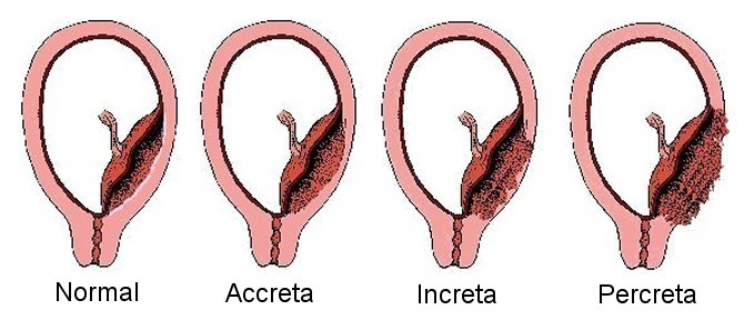
Read more...
Placental insufficiency
Failure or inability of the placenta to normally exchange nutrients, wastes,
and gases between the blood of the mother and fetus.
Giles WB, et al .,Fetal umbilical artery flow velocity waveforms and placental
resistance: pathological correlation. Br J Obstet Gynaecol. 1985
Jan;92(1):31-8. PMID: 3966988
Baschat AA, Weiner CP.Umbilical artery doppler screening for detection of the
small fetus in need of antepartum surveillance. Am J Obstet Gynecol. 2000
Jan;182(1 Pt 1):154-8.PMID: 10649171

Placenta Previa
A condition in which the placenta (including the marginal veins) partially or
completely covers the opening of the cervix .
Placental lakes (placental vascular lacunae, placental caverns, placental
venous lakes, placental sonolucencies)
Placental lakes are enlarged spaces in the placenta filled with maternal blood
called also called intervillous vascular spaces.
Placental villi
Finger-like projections of the placenta that contain fetal blood vessels. The
villi are surrounded by spaces containing maternal blood (intervillous space).
Pleural effusion
Polyhydramnios (too much amniotic fluid)
An abnormally high amount of amniotic fluid. Quantitatively an amniotic fluid
index (AFI) of 24 or more, or the largest vertical pocket of amniotic fluid
volume is 8 cm or more . Some causes of polyhydramnios include
gastrointestinal abnormalities (such as esophageal atresia and
intestinal obstruction), central nervous system abnormalities, chromosomal
abnormalities, nonimmune hydrops skeletal dysplasias diabetes twin- to -twin
transfusion. Many times no cause is found.
See also Amniotic fluid...
Ponderal Index
Postpartum
Following birth.
Postpartum blues
A common (up to 70% of women) self limiting condition occurring within a few
days of delivery. Characterized by mood lability, weeping, depression,
fatigue, anxiety, confusion, difficulty concentrating, and depersonalization
Postpartum depression
A condition (~ 10%) occurring within days to weeks following delivery and
lasting more than 2 weeks. Characterized by vegetative signs of depression,
tearfulness, anxiety, loss of interest in normal activities, guilt, inadequacy
in coping with the infant duration, thoughts of suicide. Typically requires
treatment for up to 6 months with antidepressants.
Postpartum psychosis
Uncommon condition occurring within a few days and up to 4 weeks after
delivery. Characterized by auditory hallucinations, delusions, euphoria,
grandiosity, hyperactivity, and inappropriate affect. There is a high risk of
infanticide and a high chance of developing psychosis in the future.
Treatment usually requires hospitalization.
Preeclampsia
Preeclampsia is a disease of pregnancy that affects the lining of the mother's
blood vessels causing high blood pressure, leaking of fluid from the blood
vessels, and damage to multiple organs. Preeclampsia is believed to be caused
by an abnormal placenta releasing higher than normal amounts of substances
that control the growth of blood vessels and the placenta
Pregnancy induced hypertension , PIH
Obsolete term for gestational hypertension.
Preimplantation genetic diagnosis, PGD
Testing for a specific genetic disease , such as cystic fibrosis, in an embryo prior to transferring the embryo to the uterus
.
Preimplantation genetic screening, PGS
Testing for chromosome number in an embryo prior to transferring the embryo to the uterus
. Techniques such as comparative genomic hybridization (CGH) test for
all 23 pairs of chromosomes.
Preterm
Less than 37 completed weeks' (less than 259 days) of gestation
Pseudotumor cerebri (PTC) also known as idiopathic intracranial
hypertension (IIH)
A condition of unknown cause characterized by increased intracranial pressure,
with normal cerebrospinal fluid composition, and no
abnormalities on neuroimaging. Increased pressure on
the optic nerves may cause
swelling of the optic discs (papilledema) and loss of vision. The most
common symptom of IIH is headache.
Quickening
The first movements of the fetus felt by the woman on average at 19 weeks
during the first pregnancy, and as early as 14 weeks during subsequent
pregnancies.
Red blood cell distribution width % (RDW)
The variability of red blood cell size (anisocytosis). RDW (%) = {SD of red blood cell volume (fL)/MCV (fL)} x 100
Respiratory distress syndrome (RDS, hyaline membrane disease ,HMD)
A condition of the lungs where the lungs are too stiff to expand because a
substance (surfactant) is not present to prevent the tiny air sacs in the
lungs (alveoli) from collapsing and sticking together. Damaged cells collect
in the airways and form a glassy (hyaline) membrane over the alveoli. RDS is
most likely to occur in premature infants less than 32 weeks' gestational age,
and is twice as common in boys.
Retinopathy of prematurity (ROP
The growth of abnormal blood vessels into the retina. The retina is the layer on the inside of the eye that detects light and enables you to see.
Round ligaments
The round ligaments of the uterus are two flattened bands extending from each
side of the uterus that proceed forward through a tunnel in the abdominal wall
(the inguinal canal) to the large folds of skin at the opening of the vagina
(labium majus).
Round ligament pain
Sharp pain in the lower abdomen or groin caused by spasm of the round
ligaments of the uterus. The pains usually last a few seconds and are
associated with rapid movement or rolling over during sleep.
Rupture of membranes (ROM, “breaking of the water bag” )
Breaking or tearing open of the fluid filled amniotic sac . Often described as
a "gush of fluid".
Shoulder dystocia
An average head-to-body delivery time more than 60 seconds, also defined
as "a delivery that requires additional obstetric maneuvers following failure
of gentle downward traction on the fetal head to effect delivery of the
shoulders." Shoulder dystocia is usually caused by the anterior shoulder
becoming stuck behind the mother's pubic bone.
Singleton
A pregnancy with only one fetus in the uterus.
Small for gestational age (SGA)
Weight below the 10th percentile for gestational age. Most small for
gestational age fetuses are small because of constitutional factors such as
female sex or heredity.
Smith Lemli Opitz Syndrome (SLOS)
A condition characterized by growth retardation, microcephaly,
moderate to severe intellectual disability, and malformations inluding cleft
palate, cardiac defects, underdeveloped external genitalia in males, postaxial
polydactyly, and 2-3 syndactyly of the toes. The symptoms and severity of SLOS
varies greatly in affected individuals and individuals have been
described with normal development and only minor malformations. SLOS is caused
by mutations in the DHCR7 gene. The DHCR7 gene provides instructions for
making an enzyme called 7-dehydrocholesterol reductase which is involved in
the production of cholesterol . SLOS is inherited in an autosomal recessive
manner.
Nowaczyk MJM. Smith-Lemli-Opitz Syndrome. 1998 Nov 13 [Updated 2013 Jun 20].
In: Pagon RA, Adam MP, Ardinger HH, et al., editors. GeneReviews® [Internet].
Seattle (WA): University of Washington, Seattle; 1993-2016. Available from:
http://www.ncbi.nlm.nih.gov/books/NBK1143/
Sonogram (Ultrasound)
An image or images produced by collecting sound waves reflected from
structures inside the body.
Spotting
Light vaginal bleeding.
Station
The level of the presenting part in the birth canal in relation to the ischial
spines of the pelvis. The spines represent 0 station. The presenting part is
described as being from -1 to -5 cm above the spines or +1 to+ 5 cm below the
spines. A station of + 5 cm would correspond to the presenting part at the
vaginal opening (introitus).
Stress test (Contraction stress test,CST, oxytocin contraction stress
test)
A method of testing fetal well-being and in particular the function of the
placenta under stress. The study is performed by making a graphical recording
of the fetal heart rate using an electronic monitor. The tracing is observed
for late decelerations.
Stillbirth
A fetal death that occurs during pregnancy at 20 weeks' or greater gestation.
Subchorionic hematoma
A blood clot beneath the placenta.

Succenturiate placenta
One or more accessory placental lobes connected to the main placenta
by blood vessels. There is an increased risk for postpartum hemorrhage
and infection due to retained placenta with a succenturiate placenta.
Sometimes the blood vessels that connect the lobes of the placenta cross
over or near the opening of the cervix leaving the blood vessels vulnerable to
rupture. This latter condition is called type II vasa previa
Surfactant
A substance produced in the lungs that prevents the tiny air sacs (alveoli) in
the lungs from collapsing and sticking together by reducing surface tension.
Sutures
Sutures (stitches) : Sterile, threadlike materials made of catgut, silk, or
wire used by surgeons to sew tissues together OR
Sutures : The fibrous joints between the skull bones .
Teratogen
Anything that can cause a birth defect .
Term pregnancy
The four definitions of the types of ‘term’ deliveries are:
Early Term: Between 37 weeks 0 days and 38 weeks 6 days
Full Term: Between 39 weeks 0 days and 40 weeks 6 days
Late Term: Between 41 weeks 0 days and 41 weeks 6 days
Postterm: Between 42 weeks 0 days and beyond
Tetralogy of Fallot
A birth defect of the heart consisting of :
1. Pulmonic stenosis (narrowing of the pulmonary artery).
2. A ventricular septal defect (VSD). The VSD causes cyanosis (bluish
discoloration of the skin due to lack of oxygen) by allowing blood to flow
from the right side of the heart to the left side without passing through the
lungs.
3. Malignment of the aorta so that it arises from the VSD or the right
ventricle instead of directly from the left ventricle
4. Right ventricular hypertrophy (thickening of the right heart chamber that
pumps blood to the lungs).
Thalassemia
A group of inherited blood disorders characterized by moderate to severe
anemia. Thalassemias are caused by defects in the genes that control
production of globins, the building blocks of hemoglobin (the oxygen carrying
molecule in red blood cells). The two main types of thalassemia are
alpha-thalassemia
and beta-thalassemia<
Thrombocytopenia
A lower than normal number (count) of platelets in the blood. Platelets are
cell fragments in the blood that help to form blood clots.
Titer
The concentration of an antibody in the blood.
Tocolytic
A substance that decreases uterine contractions.
Toxemia
Old name for preeclampsia
Toxoplasmosis
Toxoplasma gondii is a protozoan parasite that infects
most species of warm blooded animals and can cause the disease toxoplasmosis.
Most pregnant women who acquire the infection have no symptoms. Some may
experience malaise, low grade fever, and lymphadenopathy. Prenatally acquired
T gondii may infect the brain and retina of the fetus and can cause
chorioretinitis, intracranial calcifications, and hydrocephalus.
Transverse lie
The long axis of the fetus is perpendicular to the long axis of the mother.
The fetus is laying either on its side, back, or belly.
T-sign
On ultrasound examination the junction of two amniotic sacs forms a 90 degree
angle with the placenta. The T-sign strongly indicates that there is a single
placenta (monochorionic).
Twin peak sign, Lambda sign
On ultrasound examination the presence of a triangular projection of placental
tissue extending between two amniotic sacs. The twin peak sign strongly
indicates that there are two separate placentas (dichorionic).
Umbilical arteries
Blood vessels originating from the fetal internal iliac arteries that carry
all the oxygen depleted blood from the fetus through the umbilical cord to the
placenta.
Umbilical cord
The flexible tube that connects the fetus at the abdomen with the placenta.
Uterine contractions
Recurrent tightening and relaxation of the uterus
Uterine rupture
A tear through the entire thickness of the uterine wall.
Uterus (womb)
The pear-shaped reproductive organ in a woman's pelvis. The lower narrow part
of the uterus (the cervix) opens into the vagina.
Vacuum extraction
Traction to the infant's head through the use of a suction cup applied to the
infant's scalp for the purpose of assisting delivery.
Vaginal birth
Delivery of an infant through the birth canal (vagina).
Vaginal birth after cesarean ( VBAC )
Delivery of an infant through the birth canal in a woman who has previously
given birth by cesarean delivery.
Varicella-Zoster virus (Chickenpox, shingles)
A DNA virus of the herpes family. Infection with the virus
presents as fever followed by small papules evolving into vesicules, pustules
and crusts. The rash begins on the face and scalp then spreads to trunk. The
incubation period ranges from 10 to 21 days. The patient is contagious for 1
to 2 days before the onset of rash until all lesions are crusted. The crusts
are not infectious.
Varicella pneumonia occurs in approximately 10 % of mothers. Mortality is high
in untreated cases.
Varicella infection up to the 28th week of pregnancy has been associated with
limb hypoplasia, cicatricial lesions, psychomotor retardation, cutaneous
scars, chorioretinitis, cataracts, cortical atrophy, microcephaly,
microphthalmus, and IUGR. The risk of the syndrome is less than 2 %.
Reactivation of the varicella-zoster virus (shingles) during pregnancy does
not appear to result in intrauterine infection.
Vasa previa
Unsupported fetal blood vessels running over the cervix that are
vulnerable to bleeding. Vasa previa occurs with velamentous cord insertion and
succenturate lobes as the frees fetal vessels cross the membranes.
VATER association
An abbreviation for the combination of defects
Vertebral defects,
Anal
atresia, Tracheoesophageal fistula with Esophageal atresia, and
Radial dysplasia.
Velamentous cord insertion
Insertion of the fetal blood vessels on the membranes at the periphery (edge) instead
of directly in the middle of the placenta.
Ebbing C,et. al. Prevalence, risk factors and outcomes of velamentous and marginal cord insertions: a population-based study of 634,741 pregnancies.
PLoS One. 2013 Jul 30;8(7):e70380. PMID:
23936197
Sinkin JA, et. al., Perinatal Outcomes Associated With Isolated Velamentous Cord Insertion in Singleton and Twin Pregnancies.
J Ultrasound Med. 2017 Aug 29. PMID:
28850682
Esakoff TF, et. al., Velamentous cord insertion: is it associated with adverse perinatal outcomes?J Matern Fetal Neonatal Med. 2015 Mar;28(4):409-12. PMID:
24758363
Venous thrombosis
Formation of a blood clot inside of a vein
Ventilator
A device that mechanically assists or controls breathing continuously through
a tracheostomy or by endotracheal tube.
Vertex ( vertex presentation )
The top of the head just in front of the occipital fontanel. Vertex
presentation describes a type of cephalic presentation where the top of the
fetal head is felt through the cervix on vaginal examination.
Vernix (vernix caseosa)
The white, waxy substance that covers the skin of the fetus and newborn.
Vernix is composed of sebum (a complex mixture of fatlike compounds) and cells
that have sloughed off the fetus. Vernix is believed to act as a protective
film with anti-infective and waterproofing properties.
Very low birth weight (VLBW)
Birth weight less than 1500 grams (3 pounds 5 ounces).
Vibroacoustic stimulation (VAS)
Use of a sound emitting device placed on the maternal abdomen
to check on the well-being of the fetus
VSD ( Ventricular septal defect )
A hole in the wall that divides the large chambers of the heart (ventricles)
that pump blood.
Walking epidural
Usually refers to combined spinal-epidural anesthesia. A method of pain relief
in which anesthesia is injected into spinal fluid around the spinal cord as
well as the space around the spinal cord (epidural space). Because the dose of
the anesthesia used is much smaller than that used during a regular epidural
the muscle strength in the legs is less likely to be affected.
Wharton's jelly
A gelatin-like substance (mucoid tissue) that surrounds and protects the blood
vessels of the umbilical cord. Wharton's jelly is named after Thomas Wharton
(1614-1673) the physician and anatomist who first described it.
Womb ( uterus )
The pear shaped reproductive organ in a woman's pelvis.
X-Linked recessive trait
A trait transmitted by a gene located on the x chromosome; also called
sex-linked. Read more...
Yeast Infection
A vaginal yeast infection (also known as vulvovaginal candidiasis) is an
infection of the vagina most commonly caused by the yeast Candida albicans, a
type of fungus.
Yolk sac
A membranous structure outside of the embryo that serves as the early site for
the formation of blood.
Zidovudine ,ZDV, Retrovir, formerly called azidothymidine [AZT])
Drug used in the prevention of maternal to fetal HIV-1 Transmission
Zika virus Zika virus is a flavivirus transmitted primarily by
Aedes aegypti mosquitoes. Spread of the virus through blood transfusion and
sexual contact have been reported. The virus takes its name from the Zika
forest in Uganda where the virus was first isolated in 1947. Infection is
usually asymptomatic, and, when symptoms are present, typically results in
mild and self-limited illness with symptoms including fever, maculopapular rash, arthralgia,
and conjunctivitis. Zika virus infection has been associated with
the
development of Guillain-Barré syndrome, and in the fetus microcephaly. 1. Dick GW et. al.Zika virus. I. Isolations and
serological specificity. Trans R Soc Trop Med Hyg. 1952 Sep;46(5):509-20.
PMID: 12995440
https://trstmh.oxfordjournals.org/content/46/5/509
2. Martines RB, Bhatnagar J, Keating MK, et al. Notes from the Field: Evidence
of Zika Virus Infection in Brain and Placental Tissues from Two Congenitally
Infected Newborns and Two Fetal Losses — Brazil, 2015. MMWR Morb Mortal Wkly
Rep 2016;65(Early Release):1–2. DOI: http://dx.doi.org/10.15585/mmwr.mm6506e1er
3. Guilherme Calvet G et. al., Detection and sequencing of Zika virus
from amniotic fluid of fetuses with microcephaly in Brazil: a case study
Lancet
http://www.thelancet.com/journals/laninf/article/PIIS1473-3099(16)00095-5/abstract
Zika Virus CDC
4. Cao-Lormeau VM, et. al., Guillain-Barré Syndrome outbreak associated with Zika virus infection in French Polynesia: a case-control study.
Lancet. 2016 Apr 9;387(10027):1531-9. doi: 10.1016/S0140-6736(16)00562-6. PMID:26948433
http://www.cdc.gov/zika/index.html
Zygote ( fertilized egg )
The cell that results from fusion of a sperm and egg at fertilization. After
three divisions the zygote is called a morula.
REFERENCES
American College of Obstetricians and Gynecologists. Antepartum Fetal
Surveillance. Practice Bulletin no. 9. Washington, D.C. ACOG, 1999.
Cunningham FG. ed Williams Obstetrics, 22nd ed., New York:
McGraw-Hill.2005 Gabbe ed: Obstetrics - Normal and Problem Pregnancies, 4th ed New York, NY,
Churchill Livingstone; 2002 Jones, K.L. ed. Smith’s recognizable patterns of human malformation (5th ed.).
Philadelphia: W.B. Saunders.1997 Resnik R, ed., Maternal-Fetal Medicine, 5th ed., pp. 859–899. Philadelphia:
Saunders. Stenchever MA, Droegemueller W, eds. Comprehensive Gynecology. 4th ed. St.
Louis: Mosby, 2001 Woodward PJ ed. Diagnostic Imaging Obstetrics, 1st ed., Manitoba: Amirsys, 2005
Ogueh O, et al., Obstetric implications of low-lying placentas diagnosed in the
second trimester.
Int J Gynaecol Obstet. 2003 Oct;83(1):11-7. PMID: 14511867
|
|

